Japanese
English
- 有料閲覧
- Abstract 文献概要
- 1ページ目 Look Inside
I.はじめに
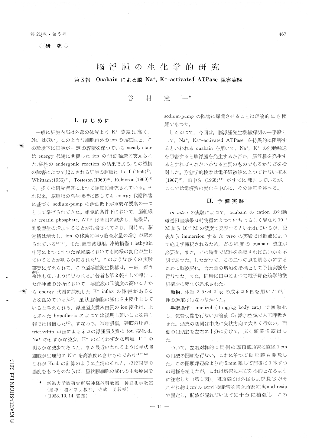
一般に細胞内部は外部の体液よりK+濃度は高く,Na+は低い。このような細胞内外のionの偏在性と,この環境下に細胞が一定の容積を保つているsteady-stateはenergy代謝に共軛したionの能動輸送に支えられた,細胞のendergonic reactionの結果である。この機構の障害によつて起こされる細胞の腫脹はLeaf (1956)1),Whittam (1956)2),Tosteson (1960)3),Robinson (1960)4)ら,多くの研究者達によつて詳細に研究されている。それ以来,脳腫脹の発生機構に関してもenergy代謝障害に基づくsodium-pumpの活動低下が重要な要素の一つとして挙げられてきた。嫌気的条件下において,脳組織のcreatin phosphate, ATPは著明に減少し,無機P,乳酸産生の増加することが報告されており,同時に,脳容積は増大し,ionの移動に伴う脳含水量の増加が認められている5)〜7)。また,超音波照射,凍結損傷triethyltin中毒によつて作つた浮腫脳においても同様の変化が生じていることが明らかにされた8)。このような多くの実験事実に支えられて,この脳浮腫発生機構は,一応,疑う余地もないように思われる。著者も第2報として報告した浮腫液の分析において,浮腫液のK濃度の高いことからenergy代謝に共軛したK+inflaxの障害があることを認めているが9),星状膠細胞の膨化を主変化としていると考えられる,浮腫脳皮質灰白質のion変化は,上に述べたhypothesisによつては説明し難いことを第1報では指摘した10)。すなわち,凍結損傷,硬膜外圧迫,triethyitin中毒によるネコの浮腫脳皮質のion変化は,Na+のわずかな減少,K+のごくわずかな増加,Cl—の明らかな減少であつた。また最近いわれるように星状膠細胞が生理的にNa+を高濃度に含むものであり11)〜3),これがKochの計算のように血清のそれと,ほぼ同等の濃度をもつものならば,星状膠細胞の膨化の主要原因をsodium-pumpの障害に帰着させることは理論的にも困難であつた。
したがつて,今回は,脳浮腫発生機構解明の一手段として,Na+,Ka+—activated ATPaseを特異的に阻害するといわれるouabainを用いて,Na+,K+の能動輸送を阻害すると脳浮腫を発生するか否か,脳浮腫を発生するとすればそれがいかなる性質のものであるかなどを検討した。形態学的検索は電子顕微鏡によつて行ない植木(1967)8),田中ら(1968)14)がすでに報告しているが,ここでは電解質の変化を中心に,その詳細を述べる。
In the first report of this series, it was indicated that an extreme difficulty existed in explaining the move of electrolytes in edematous gray matter by the theory of sodium-pump impairment. In order to ascertain whether or not the primary inhibition of the membrane ATPase was responsible for pro-ducing a state of brain edema, present experiment was performed using ouabain as a Na+, K+-activated ATPase inhibitor.
To determine the ouabain concentration and the time of immersion enough to produce edematous change, the changes in water content and electroen-cephalogram were investigated in the preliminary experiments using 9 adult cats. For this purpose experimental animal was lightly anesthetized with Nembutal, the trachea was cannulated low in the neck and artificial respiration was used after respi-ratory paralysis was induced by intramuscular in-jection of Amelizol. Two burr holes of 10 mm in diameter were opened through the parietal bones, then brain surface was carefully exposed and kept moist with physiological saline. Each two electrodes were fixed anteriorly and posteriorly at a distance of 5 mm from the edge of each hole, being exactly symmetric to each other. Bipolar EEG recording was led from each cortical surface before and after the immersion. The electron microscopic examination carried out by other investigator in the laboratory on the material which showed a spongy state in light microscopy revealed the swelling of cell pro-cesses, mainly of neuronal element, and the swelling was confined to synaptic region, particularly to the post synaptic elements. Even in the severely af-fected lesion, perikarya of astrocytes and pericapillary end-feet showed no evidence of swelling.
Based on these data, following experiment was carried out with 9 adult cats immersed with 10-3M ouabain solution for 40 minutes before sacrifice. Determination of water, electrolytes and protein contents and polyacrylamide gel electrophoretic ana-lysis of protein were carried out on the edematous gray matter dissected from the cortex immersed with ouabain solution by the same technique which were described in the previous reports of this series.
The results were : ( 1 ) From ouabain side, EEG changes of rhythmic rapid rhythms were recorded within 1 minute after immersion, then slow com-ponents progressively increased among those rapid rhythms. Usually after 30 minutes of ouabain im-mersion, EEG recording showed flat pattern. Also, rhythmic rapid rhythms gradually appeared in the control side, and that lasted throughout the experi-ment. Neither voltage accentuation nor spikes were observed. ( 2 ) Slight increase in water, marked increase in sodium and negligible change in chloride contents were revealed. Nervertheless total protein was in normal range and the soluble protein showed distinct increase without remarkable changes in poly-acrylamide gel electrophoretograms. ( 3 ) These changes, particularly those in electrolytes movement, differ greatly from previous findings observed in the edematous brain produced by epidural compression and cold injury. It was therefore highly probable that the cation transport system across cell membrane was responsible for cell swelling in neuronal rather than glial elements.

Copyright © 1969, Igaku-Shoin Ltd. All rights reserved.


