Japanese
English
- 有料閲覧
- Abstract 文献概要
- 1ページ目 Look Inside
I.はじめに
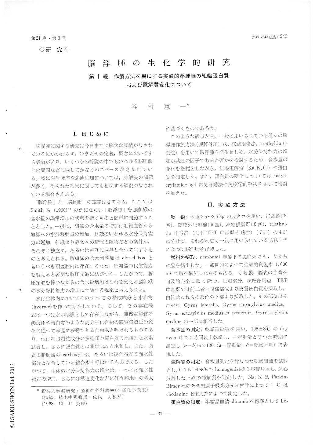
脳浮腫に関する研究は今日までに膨大な集債がなされているにかかわらず,いまだその定義,概念においてすら議論があり,いくつかの総説の中でもいわゆる脳腫脹との異同などに関してかなりのスペースがさかれている。特に発生機序や病態生理については,未解決の問題が多く,得られた結果に対しても相反する解釈がなされている場合さえある。
「脳浮腫」と「脳腫脹」の定義はさておき,ここではSmithら(1960)1)の例にならい「脳浮腫」を脳組織の含水量の異常増加の状態を指すものと簡単に制約することとした。一般に,組織の含水量の増加は毛細血管から組織への水分移動量の増加,組織のいわゆる水分保持能力の増加,組織より静脈への環流の阻害などの条件が,それぞれ独立に,あるいは相互に関与し合って生ずるものと考えられる。脳組織の含水量増加はclosed boxともいうべき頭蓋腔内に存在するため,脳組織の代償能力を越えると著明な脳圧亢進に結びつく。したがつて,脳圧亢進を伴いながらの含水量増加はこれを支える脳組織の水分保持能力の増加に付随する現象と考えられる。
In order to find common phenomena in edematous brain, the following methods were employed in pro-ducing cerebral edema in 28 adult cats, i. e., epidural compression, cold injury and triethyltin intoxication, and hydrophilic solutes of brain tissue such as elec-trolytes and proteins were analysed.
For all experimental groups, white matter and cerebral gray matter of the same region were dis-sected and water content of brain tissue was deter-mined with drying method. The dried samples were extracted with 0.1 N HNO3, and sodium and potas-sium content was determined with atomic absorption spectrophotometry and Lowry's rhodanine colorimetry for chloride. Total protein and water soluble protein extracted from the tissue samples were determined by Lowry's technique. Polyacrylamide gel electro-phoretic analysis of the soluble protein was carried out according to the methods described by Ornstein and Davis, with a variation that the concentration of acrylamide in small pore gell was 9.5 per cent, to make clearer the fractionation of proteins relatively smaller than plasma albumin. To examine the presence of albumin and other plasma proteins in the edematous brain, immunoelectro-phoretic analysis and Ouchlerlony's double diffusion test were made on the soluble protein using the antiserum against cat serum protein. Whole blood content of the tissue samples was estimated by the method of Gordon and Numberger.
Results were : (1) An increase in the water con-tent of the brain characteristically occurred in white matter to a greater degree than in gray matter in all experimental groups.
(2) The changes in the electrolytes content of gray matter were peculiar, i.e., the minimum or negligible change in sodium and slight increase in potassium and moderate decrease in chloride (10-20 per cent). These phenomena may be explained as are the result of narrowing of extracellular space due to the swelling of astrocytes. By contrast, in edematous white matter, increase in sodium and chlo-ride and decrease in potassium were common pheno-mena, which agrees with the fact reported by others. Especially, the chloride content well paralleled with the increase of water content. These findings clearly indicated that the location of edema fluid in the white matter was in the extracellular space.
(3) In general, changes in protein content of edematous brain tended to separate into two groups, that of TET poisoning and that of other. Namely, in the TET poisoning group, total protein content of cerebral white matter and cortex were both within normal range, but soluble protein in gray matter was elevated. In other group, gray matter showeddecrease in total protein content and increase in soluble protein content, although white matter showed increase both in the total and soluble protein content.
(4) About twenty bands of soluble protein were distinguished on polyacrylamide gel electrophoresis in all groups and the profile of electrophoretograms showed characteristics for each experimental group. Most remarkable and common changes in electro-phoretograms among brain edema groups, except for the TET poisoning, were the intensive increase in albumin fraction identified with serum albumin using Ouchterlony's technique.
Another remarkable and common change in the electrophoretogram was the relative increase in pro-tein fraction which migrated more rapidly than albumin. This was reflected in the elevation of F-B protein ratio (the densitometric scan of electro-phoretogram was divided into three zones; F-zone containing protein migrated faster than albumin, Albumin zone and B-zone containing protein mi-grated slowlier than albumin). Considering the changes in protein content mentioned above, these changes in electrophoretogram of the gray matter can be explained to be the result of solubilizing processes of protein composing the subcellular units, and in white matter these may he responsible for this elevation.
(5) In immunoelectrophoretic analysis of TET poisoning and normal brain tissus no precipitation lines could be detected. In the other group of brain edema, an albumin precipitation line was usually detected. Occasionally in some of the edematous white matter of cold injury group, several globulin precipitation lines were detected but they were distinctly fewer than those of the serum ob-tained from the same animal.

Copyright © 1969, Igaku-Shoin Ltd. All rights reserved.


