Japanese
English
- 有料閲覧
- Abstract 文献概要
- 1ページ目 Look Inside
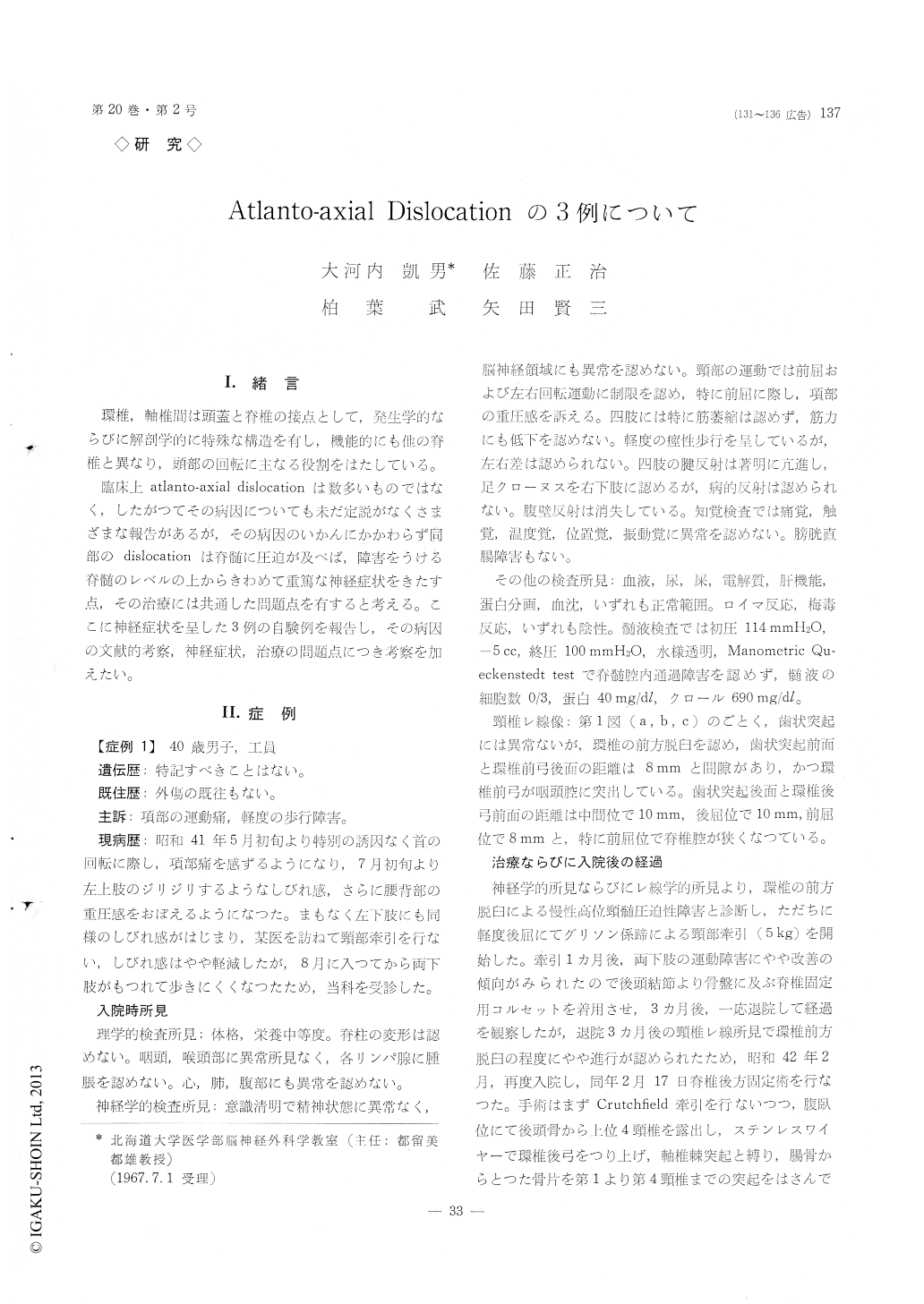
I.緒言
環椎,軸椎間は頭蓋と脊椎の接点として,発生学的ならびに解剖学的に特殊な構造を有し,機能的にも他の脊椎と異なり,頭部の回転に主なる役割をはたしている。
臨床上atlanto-axial dislocationは数多いものではなく,したがつてその病因についても未だ定説がなくさまざまな報告があるが,その病因のいかんにかかわらず同部のdislocationは脊髄に圧迫が及べば,障害をうける脊髄のレベルの上からきわめて重篤な神経症状をきたす点,その治療には共通した問題点を有すると考える。ここに神経症状を呈した3例の自験例を報告し,その病因の文献的考察,神経症状,治療の問題点につき考察を加えたい。
3 cases of atlanto-axial dislocation with neuro-logical manifestations are reported.
Case 1 : 40 year-old male came in with progressive gait disturbance and neck pain of 3 months duration. Neurological examination on admission revealed mild spasticity and hyperactive deep tendon reflexes in all four extermities, but sensory examination was completely negative. X-ray examination showed anterior dislocation of the atlas which was most marked when the neck was flexed (fig. 1). The pa-tient was first treated with neck traction for ap-proximately one month. As neither of the clinical signs nor the X-ray evidence of the dislocation was improved, posterior fusion of the C1 to C4 together with occipital bone was performed. For months after the operation, the fusion was completed as shown on fig. 2, but the neurological signs remained the same.
Case 2: 20 year-old male admitted with progres-sive gait disturbance of several months duration and epileptic seizures since his childhood. On admission, the patient showed mild spasticity of all 4 extrem-ities which was more marked on the left side. There were no demonstrable sensory disturbances. X-ray examination revealed atlanto-axial dislocation withos odontoideum. The dislocation was most exagger-ated on flexed position as shown on fig. 3. Because of the marked instability of the joint, posterior fusion of C1 to C4 together with occipital bone was per-formed and the result was satisfactory (fig. 5).
Neurological symptoms and signs remained the same except for disappearance of the epileptic seizure attacks by using anticonvulsants.
Case 3: 58 year-old man, admitted after banging on his occiput by falling on the ground. All of the extremities were completely paralyzed right after the fall, but they began to move approximately 10 minutes after the fall starting from left arm. Al-though the patient showed marked spastic quadri-paresis, there was no sensory disturbances of any kind, The size of the head was markedly enlarged and also showed a basilar impression as shown fig. 6. Neck X-ray revealed a marked degree of atlanto-axial dislocation with isolated os odontoideum (fig. 7). The patient refused operation and after neck traction. of 6 weeks' duration, motor weakness was markedly improved.
Neurologically, it is very interesting that these cases show only mild spasticity without any sensory disturbances having such a marked dislocation of atlanto-axial joint. This is probably due to the fair-ly large size of the bony canal at this level com-paring the size of the spinal cord. However, as shown on the Case 3, these patients always have potential danger of upper cervical cord injury even by a fairly mild force of hyperflexion. Therefore, we feel it is necessary to fuse upper cervical ver-tebra in these cases.
In cases 2 and 3, whether the detached tips of the odontoid processes are so called un-united os odontoideum or they are ununited fractured odontoid process is not completely clear. However, from the history that they have never had a head or neck injury and that they have other congenital abnor-malities, we assume that these are also congenital un-united os odontoideum.
Copyright © 1968, Igaku-Shoin Ltd. All rights reserved.