Japanese
English
- 有料閲覧
- Abstract 文献概要
- 1ページ目 Look Inside
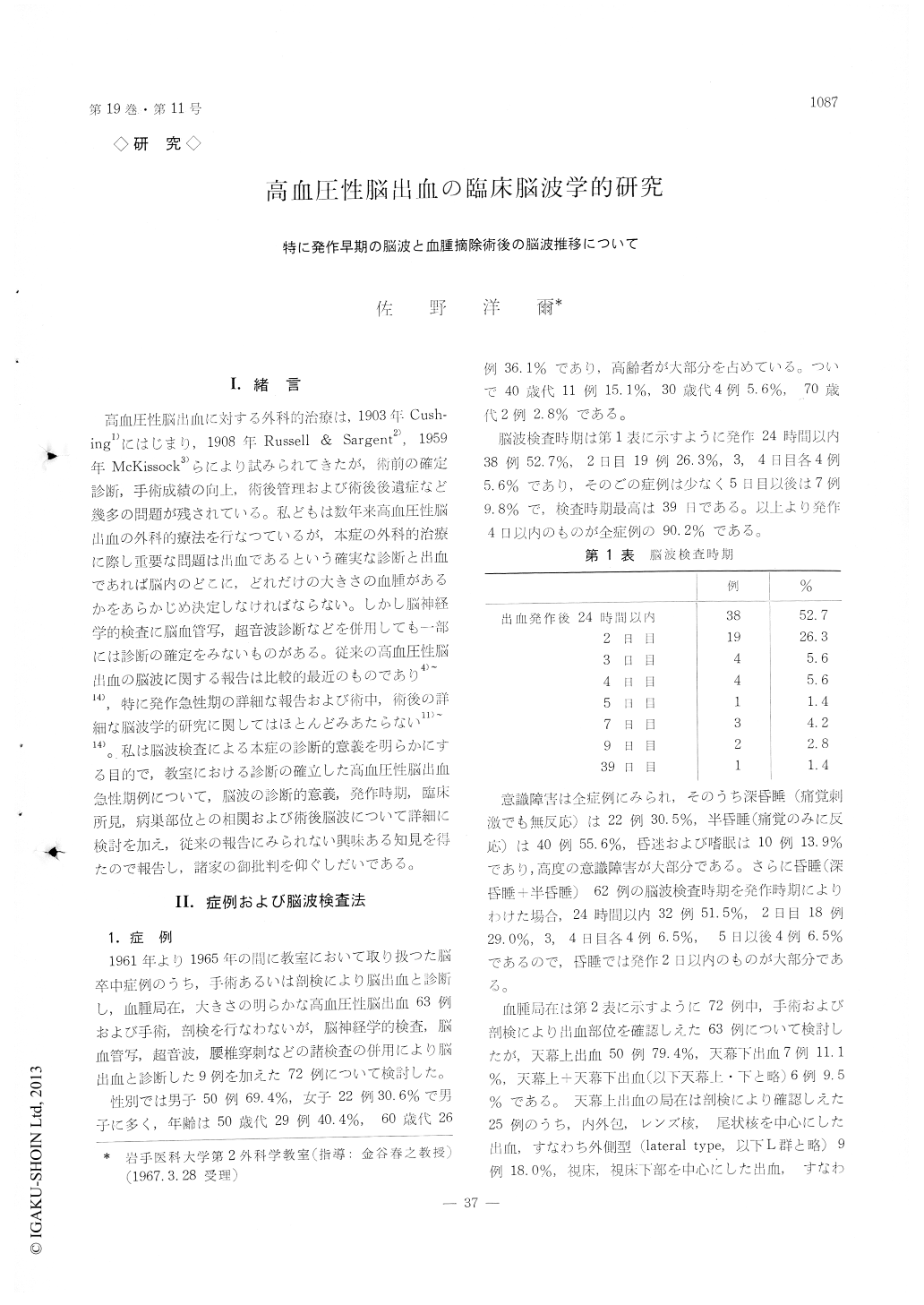
I.緒言
高血圧性脳出血に対する外科的治療は,1903年Cush—ing1)にはじまり,1908年Russell&Sargent2),1959年McKissock3)らにより試みられてきたが,術前の確定診断,手術成績の向上,術後管理および術後後遺症など幾多の問題が残されている。私どもは数年来高血圧性脳出血の外科的療法を行なつているが,本症の外科的治療に際し重要な問題は出血であるという確実な診断と出血であれば脳内のどこに,どれだけの大きさの血腫があるかをあらかじめ決定しなげればならない。しかし脳神経学的検査に脳血管写,超音波診断などを併用しても一部には診断の確定をみないものがある。従来の高血圧性脳出血の脳波に関する報告は比較的最近のものであり4)〜14),特に発作急性期の詳細な報告および術中,術後の詳細な脳波学的研究に関してはほとんどみあたらない11)〜14)。私は脳波検査による本症の診断的意義を明らかにする目的で,教室における診断の確立した高血圧性脳出血急性期例について,脳波の診断的意義,発作時期,臨床所見,病巣部位との相関および術後脳波について詳細に検討を加え,従来の報告にみられない興味ある知見を得たので報告し,諸家の御批判を仰ぐしだいである。
We have been performing the surgical treatment on the cases of hypertensive intracerebral hemor-rhage these several years. First and the most im-portant is to ensure that the patient has hemorrhage, and then the site and volume of the hematoma should be defined.
On seventy-two cases thus diagnosed in our clinic between 1961 and 1965, diagnostic significance of EEG, time of stroke, clinical findings and the post-operative EEG were studied.
Results were summarized as follows:
1) The EEG of the patients was consisted mainly of slow -a wave and θ- wave, while δ- wave which has currently been said to be characteristic in this disease, and the focal sign were both seen but a few.
2) Asymmetry in EEG appeared and increased gradually after stroke along with the regular slow -a wave from the opposite site of hematoma.
3) Worse the conscious disturbance was, more the irregular slow -α waves were seen.
4) As intracranial pressure became higher, fast-wave, spike- and sharp- wave and the irregular slow wave were increased and in many cases the EEG pattern was asymmetric.
5) Patients who made little improvement postopera-tively were found to have shown more θ- waves in his preoperative EEG.
6) Localization of EEG clearly indicated the hema-toma site. In supratentorial hemorrhage, fast-wave, spike- and sharp -wave and asymmetric pattern were obtained as well as the irregular slow -α wave from the hematoma site. Subtentorial hemorrhage, on the other hand, was suggested by the symmetric am-plitude of the left and right sides and the regular slow -α wave was obtained from the hematoma site.
7) Asymmetric pattern was frequent in the EEG of thore with hematoma volume of 51 gramm or more.
8) Fast-wave and spike- and sharp-wave were often found in EEG of a patient with ventricular hemor-rhage and neither was recorded form the case with no such hemorrhage but δ- wave and asymmetric pattern instead.
9) Slow waves vanished soon after operation when the patient's postoperative condition got better. At the same time there was an appearance of regular slow-wave and the subsequent EEG would pass into the normal pattern, correlating to his postoperative improvement.
Copyright © 1967, Igaku-Shoin Ltd. All rights reserved.