Japanese
English
- 有料閲覧
- Abstract 文献概要
- 1ページ目 Look Inside
I.はじめに
内水頭症が成立つには髄液の生成,吸収,あるいはその流れの異常が関係することはいうまでもない。髄液の生成,吸収については病理形態学的に把握することは容易なことではないが,後者には流れの異常をひき起こす重大な原因である流通部位の狭窄ないし閉塞性病変があり,これは形態学的につかみうるのである。特に生理的に通路の狭い部分すなわち室間孔,中脳導水管,脳室と髄膜腔との移行部であるMagendie孔,Luschka孔などには閉塞性病変が往々にしてみられる。その閉塞性病変の発生にはその管を形成する組織自身の病変によつても,また近接組織病変の波及,あるいはその病変の圧迫によつてもひき起こされうる。病変としては形成異常,炎症性病変あるいはその後遺症,腫瘍などが考えられる。ここに特異な,各々異なつた原因によるMonro孔,あるいは中脳導水管の閉塞性病変によりひき起こされたと考えられる内水頭症の2例を報告する。
Case 1
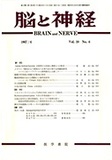
A 57 year old female had had hypertension. She was admitted to the hospital with symptoms of pal-pitation, dyspnea and arrythmia perpetua. She expired suddenly. No neurological symptom was observed. Autopsy findings revealed haemosiderosis of dura mater, sclerosis of cerebral arteries, slight old subarach-noidal haemorrhages on bilateral frontal and left occipital lobes. Cut sections showed left foramen Monroi to be occluded by thin membranous tissue adhering to plexus chorioides and marked dilatation of left lateral ventricle. There were rice sized ence-phalomalacic cysts in right putamen, bilateral thalami and old haemorrhage in right internal capsule. The membranous tissue was composed of glial tissue to be lined with ependymal cells. Paraventricular tissue of left anterior portion of third ventricle and of anterior horn of left lateral ventricle showed glial proliferation containing large glial cells and ependymal cells. There were microscopic villous projections of glial cells on ependymal lining of third and bilateral lateral ventri-cles. No inflammatory cell infiltration was seen. These chahges were considered to be malformed.
Case 2
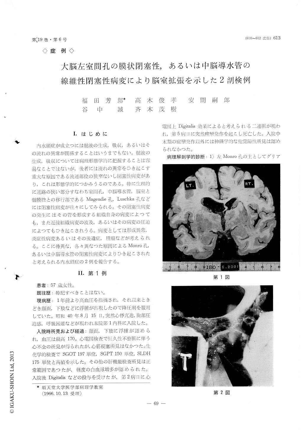
A 26 year old male had had osteomyelitis and sepsis at the age of 14 years. He had fever, chill, headache, and vomiting for 2 months. At the admission, menin-gism and ptosis of right palpebra were observed. Spinal fluid; clear, pressure 230~150, cell count 660/3, protein 240mg/dl Nonne-Apelt (++), pandy (+++),trypto-phan (-), bacteriological examination (-). He expired on 18th hospital day. Autopsy findings revealed pineal body mostly to be replaced by fibrous connective tissue extending to posterior portion of mesencephalon. Aquaeductus Sylvii was occluded by the fibrosis and the surrounding proliferated glial cells containing ependymal cells. There was marked dilata-tion of third and bilateral lateral ventricles. Lympho-cytic infiltration was observed in pineal body as well as in the surrounding tissue, subependymal perivas-cular areas and subarachnoidal space. The case was considered to be non-specific chronic inflammation of pineal body area causing occlusion of aqueductus Sylvii.
Copyright © 1967, Igaku-Shoin Ltd. All rights reserved.