Japanese
English
- 有料閲覧
- Abstract 文献概要
- 1ページ目 Look Inside
I.緒言
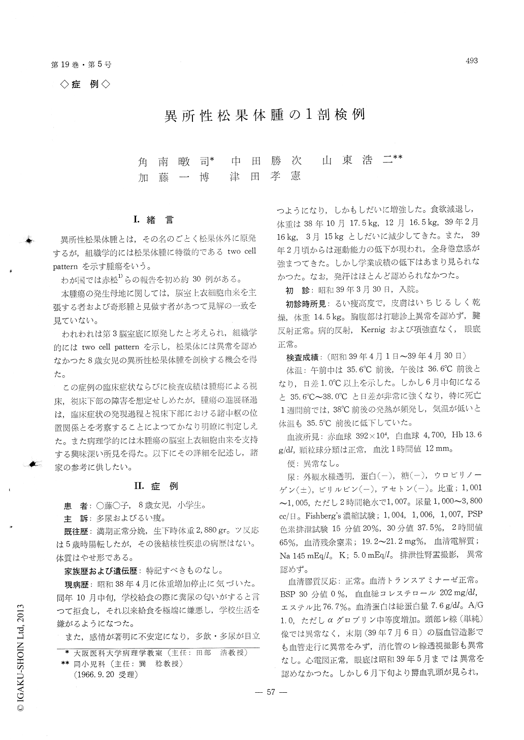
異所性松果体腫とは,その名のごとく松果体外に原発するが,組織学的には松果体腫に特徴的であるtwo cellpatternを示す腫瘍をいう。
わが国では赤松1)らの報告を初め約30例がある。
The patient is a 8 years old girl. She has no parti-cular past illness except for measles and munps at her age of. 3. The initial symptom which appeared 11 month prior to her administration to the hospital was loss of her weight. The heterosmia, polyuria, anorexia and emotional lability were followed.
She admitted to the hospital at March 1964. Her body temperature was labil. Vomitus and papilledema occured and increased gradually. No sign of the sexual precox was seen. Laboratory examinations showed marked decrease in the function of both the pituitary gland and the target endocrine glands, such as adrenals and thyloid gland. Blood sugar was markedly labile, ranged 260mg/dl to 30mg/dl, at terminal stage. She died on July 7. 1964.
The entire course of her illness since onset of the initial symptom, was 1 year and 3 month.
At autopsy, the tumor, which is about a walnut size and dark red in color, was found in the 3rd ventricle.
The tumor occupied the 3rd ventricle lumen and opressed laterally the thalamus and hypothalamus, but no infiltration of the tumor cell to the 3rd ventricular wall was found. The pineal body was in the original site and appeared to be normal bath macroscopically and microscopically.
The lateral ventricles of both sides were dilatated, but no abnormality was found in their wall and in the other parts of the brain. The pituitary gland appeared macroscopically to be normal, but histological sections of the pituitary revealed that the posterior lobe was replaced by the tumor cell and the anterior lobe was markedly atrophy. The tumors were characterized by presence of the so-called two cell patern, which con-sisted of large tumor cell and small round cell. Some of the large tumor cells had a cone-shape-profection of the cytoplasma and showed a rosette like structure. In some place the tumor cells arranged in a lineal fashion which is very similar to the arrangement of the epen-dymal lining.
A small number of tumor cells was found at the subependymal layer of the aqueduct Sylvi.
The author considered that the present tumor was a so-called ectopic pinealoma and must have arised from the ependymal cells at the bottom of the 3rd ventricle because of following reasons; 1) the pineal body was intact, 2) the histological finding of the tumor was the same as the pinealoma described in the litarature, 3) the histological structure of the tumor resembled to that of the ependymal cell in some respects.

Copyright © 1967, Igaku-Shoin Ltd. All rights reserved.


