Japanese
English
- 有料閲覧
- Abstract 文献概要
- 1ページ目 Look Inside
I.はじめに
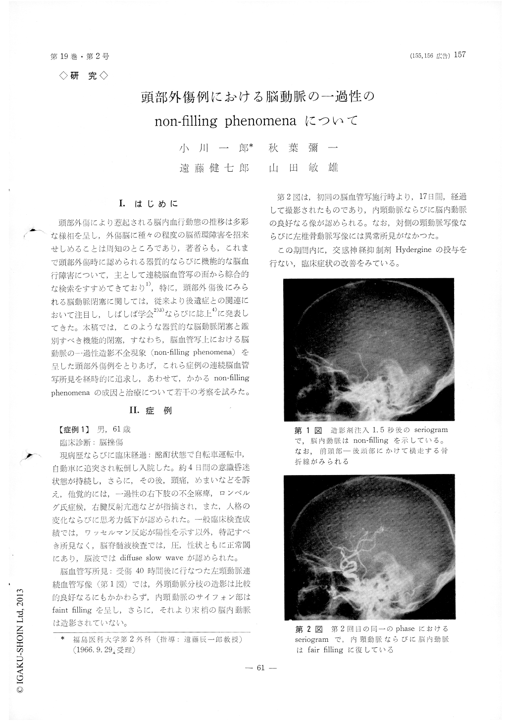
頭部外傷により惹起される脳内血行動態の推移は多彩な様相を呈し,外傷脳に種々の程度の脳循環障害を招来せしめることは周知のところであり,著者らも,これまで頭部外傷時に認められる器質的ならびに機能的な脳血行障害について,主として連続脳血管写の面から綜合的な検索をすすめてきており1),特に,頭部外傷後にみられる脳動脈閉塞に関しては,従来より後遺症との関連において注目し,しばしば学会2)3)ならびに誌上4)に発表してきた。本稿では,このような器質的な脳動脈閉塞と鑑別すべき機能的閉塞,すなわち,脳血管写上における脳動脈の一過性造影不全現象(non-filling phenomena)を呈した頭部外傷例をとりあげ,これら症例の連続脳血管写所見を経時的に追求し,あわせて,かかるnon-filling phenomenaの成因と治療について若干の考察を試みた。
Non-filling of the cerebral arteries with contrast material during serial cerebral angiography has been demonstrated in four patients with head injury.
1) The degrees of the injury of these cases, all male, ranging from 18 to 64 in age, are rather sev-er; cerebral laceretion (2), intracranial hematoma (1) and suspected skull base fracture (1).
2) The affected sites of the cerebral arteries are disclosed at the distal portion of the carotid siphon (2) and the anterior cerebral artery system (2), in a case of the latter, local non-filling of the per-icallosal segment is observed.
3) These non-filling phenomena are certified at the first serial angiography performed within three weeks after the head in jury and failed to exist on the next seriogram obtained 2-6 weeks there-after.
4) During the term between the first and the second angiography, some medical treatments of ATP, cytochrome C, manitol and stellate ganglion block are applied to these cases.
5) Moreover, systematic administration of Hyd-ergine, one of the powerful sympathetic inhibiting agents was tried to all of these cases with fairly good results in both clinical and angiographic find-ings.
6) As etiologic considerations, these non-filling phenomena are presumed to be owing to the vaso-spastic alteration in three cases, on the other hand, to the compression of the vascular segment by cer-ebral edema in a case with intracranial hematom.

Copyright © 1967, Igaku-Shoin Ltd. All rights reserved.


