Japanese
English
- 有料閲覧
- Abstract 文献概要
- 1ページ目 Look Inside
はじめに
最近,脳卒中例の脳血管撮影に於て,early ven—ous fillingやcapillary blushに関して興味ある知見が報告されており3,14〜16),実験的所見と併せてその発現機序が種々論じられている5〜9)。一方外傷脳におけるearly venous fillingやcapillary blushの記載は少なく,2,3の臨床的報告がみられるに過ぎない1,3,7)。
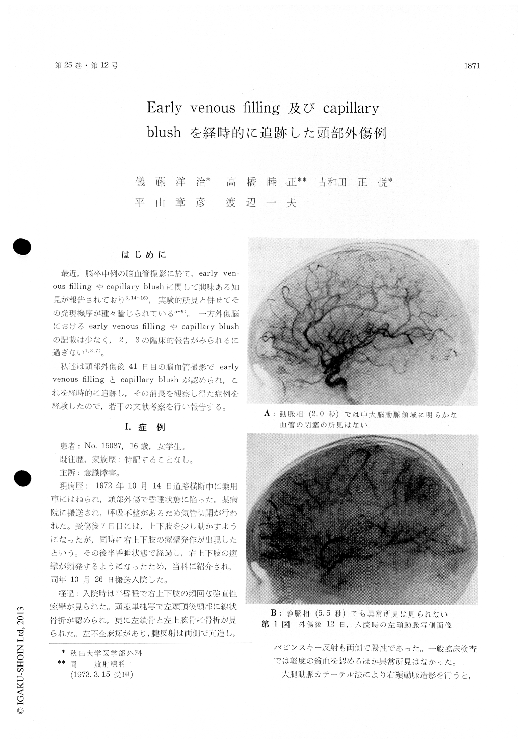
私達は頭部外傷後41日目の脳血血管撮影でearlyvenous fillingとcapillary blushが認められ,これを経時的に追跡し,その消長を観察し得た症例を経験したので,若干この文献考察を行い報告する。
An early venous filling and a capillary blush have been demonstrated 41 days after head injury. Follow-up studies of these phenomena have been reported herein in detail with discussion.
A 16-year-old school girl was admitted two weeks after a car accident on October 26, 1972. Right carotid angiography revealed a contralateral dis-placement of the anterior cerebral artery and an elevation of the right middle cerebral artery, suggesting a mass lesion in the right temporal region. Subdural and intracerebral hematomas in the right temporal area were evacuated shortly after the admission.
Bilateral serial carotid angiographies done 41 days after the head injury demonstrated an earlyvenous filling and a capillary blush in the parietal region of both sides far from the operated area. Brain scanning (99mTc) revealed also a high activity area in bilateral region.
Follow-up angiography carried out 53 days after the episode showed the same findings as the second angiogram. The early venous filling and the capil-lary blush dissapeared on the fourth angiogram performed 65 days after the accident.

Copyright © 1973, Igaku-Shoin Ltd. All rights reserved.


