Japanese
English
- 有料閲覧
- Abstract 文献概要
- 1ページ目 Look Inside
I.緒言
トリ類の視覚路,眼筋支配中枢などに関してはBello—nci1)(1888), Bradis3) (1895),Edinger,et al.9)(1899),Ris23)(1899),Rendahl22)(1924),Kunkel19)(1928),Hu—ber,et a1.14)(1929),Koikegami17)(1933),Yoshida30)(1953),Cowan,et al.5)(1961)などの報告があり,これらはいずれも正常例または実験例に基づくものである。これに対して,先天性異常例を扱った報告はBrouwer4)(1918),Mikami,et al.21)(1953)などきわめて少ない。しかし先天性異常例は発生学的および形態学的研究には重要なものであつて,実験では得られない大きな意義がある。
著者は東京大学医学部解剖学教室中井準之助教授の御好意により眼に異常の認められる貴重なニワトリ胎児を入手する機会を得たので,その所見のうち主として視覚路について報告し,視覚系の発生学的研究の一資料としたい。
A twelve-clay old chick embryo with its left eye malformed was fixed with the Bouin's fluid, embed-ded in paraffin, sectioned at 10 p. and stained after Bobian and Kluver-Barrera. The findings are summerized as follows :
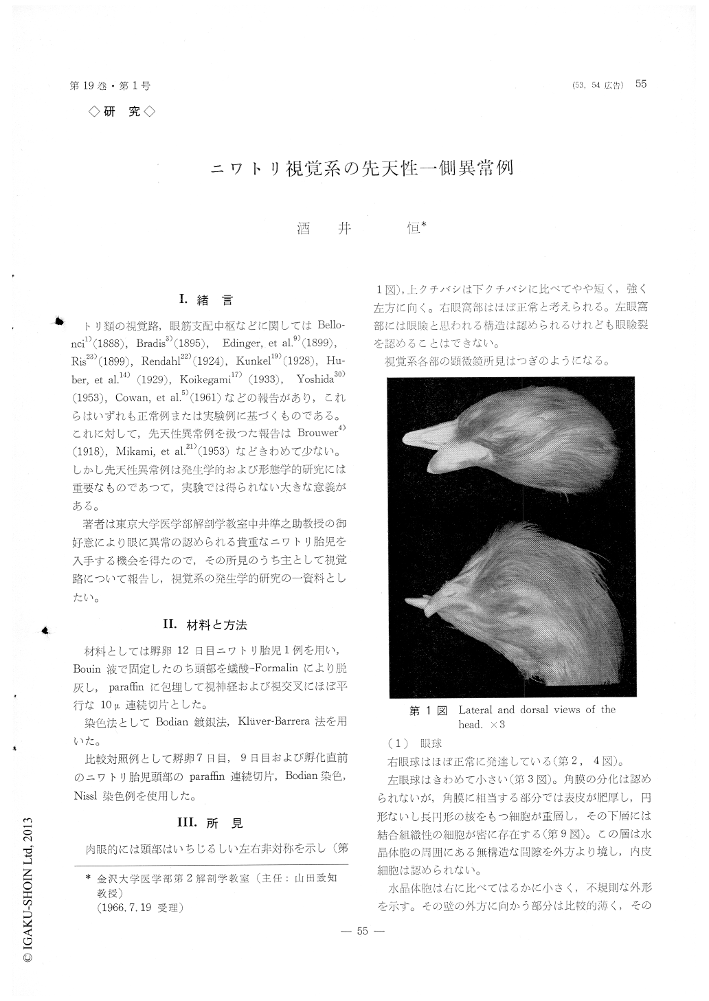
The left eyeball shows a strong hypoplasia (Fig. 3) in comparison with the right eye which appears to be normal (Figs. 2, 4). The optic cup of the left side is malformed (Figs. 11, 13), as it represents a closed sphere which is lined with the pigment layer all over, except the entrance of the optic nerve. The lens is situated in front and outside the optic cup (Fig. 11). The ocular muscles are also strongly un-der-developed.
The left optic nerve (Figs. 5, 13), which is stron-gly reduced, reaches as far as the lateral geniculate A twelve-clay old chick embryo with its left eye malformed was fixed with the Bouin's fluid, embed-ded in paraffin, sectioned at 10 p. and stained after Bobian and Kluver-Barrera. The findings are summerized as follows :
The left eyeball shows a strong hypoplasia (Fig. 3) in comparison with the right eye which appears to be normal (Figs. 2, 4). The optic cup of the left side is malformed (Figs. 11, 13), as it represents a closed sphere which is lined with the pigment layer all over, except the entrance of the optic nerve. The lens is situated in front and outside the optic cup (Fig. 11). The ocular muscles are also strongly un-der-developed.
The left optic nerve (Figs. 5, 13), which is stron-gly reduced, reaches as far as the lateral geniculate nucleus of the same side, along with the normal optic fibres of the contralateral origin. The right optic tract is not formed at all, and the lateral geniculate nucleus of this side is only poorly developed (Fig. 7). The right optic tectum is also under-developed in such a way that its 1 st layer (Ris, 1899) is lackin gand that the 2nd to 5th layers are thinner than the left side. Especially thin are the layers 2a, 3a and 5a (Fig. 8).
The left oculomotor nerve is slightly more slen der than the right nerve (Fig. 17). As regards the nucleus, the dorsolateral and the dorsomedial nuclei of the left side are under-developed in comparison with the right nuclei. The nucleus of Edinger-West-phal and the nuclei arciformis and paraccessorius (Koikegami, 1933) are under-developed on the left side (Fig. 15). The abducent nerve as well as its nucleus of the left side is under-developed also.
The finding that the lens vesicle discontinuos its differentiation as the retina remains insulated leads to the following conclusion. For the later development and differentiation of the lens vesicle, it appears that a continued influence on it of the inner wall of the retina is necessary.
The development of the lateral geniculate nucleus is greatly affected by the optic fibres as well as the supraoptic commissural fibres.
Copyright © 1967, Igaku-Shoin Ltd. All rights reserved.