Japanese
English
- 有料閲覧
- Abstract 文献概要
- 1ページ目 Look Inside
I.はじめに
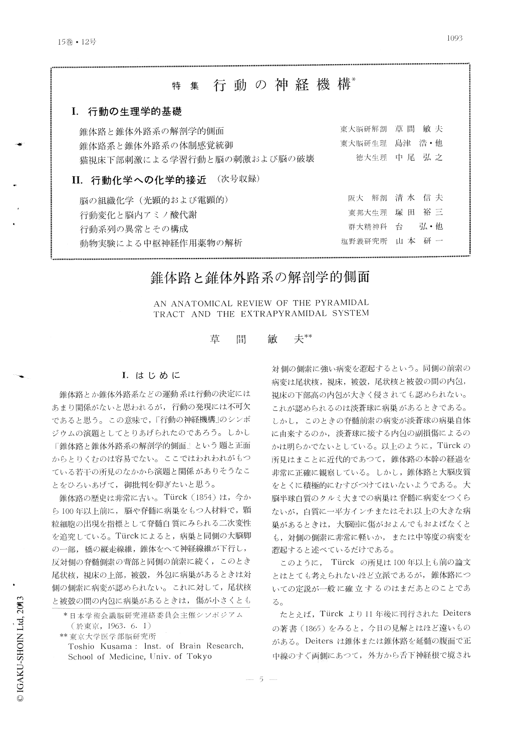
錐体路とか錐体外路系などの運動系は行動の決定にはあまり関係がないと思われるが,行動の発現には不可欠であると思う。この意味で,「行動の神経機構」のシンポジウムの演題としてとりあげられたのであろう。しかし「錐体路と錐体外路系の解剖学的側面」という題と正面からとりくむのは容易でない。ここではわれわれがもつている若干の所見のなかから演題と関係がありそうなことをひろいあげて,御批判を仰ぎたいと思う。
錐体路の歴史は非常に古い。Türck (1854)は,今から100年以上前に,脳や脊髄に病巣をもつ人材料で,顆粒細胞の出現を指標として脊髄白質にみられる二次変性を追究している。Türckによると,病巣と同側の大脳脚の一部,橋の縦走線維,錐体をへて神経線維が下行し,反対側の脊髄側索の背部と同側の前索に続く,このとき尾状核,視床の上部,被殻,外包に病巣があるときは対側の側索に病変が認められない。これに対して,尾状核と被殻の間の内包に病巣があるときは,傷が小さくとも対側の側索に強い病変を惹起するという。同側の前索の病変は尾状核,視床,被殻,尾状核と被殻の間の内包,視床の下部高の内包が大きく侵されても認められない。これが認められるのは淡蒼球に病巣があるときである。しかし,このとぎの脊髄前索の病変が淡蒼球の病巣自体に由来するのか,淡蒼球に接する内包の副損傷によるのかは明らかでないとしている。
Definition of the pyramidal tract and the extrapyramidal system was discussed on lite-ratures and on our findings with the Nauta method. Our findings cited are as follows.
1) The cortical sensory leg area of cat, the medial part of the posterior sigmoid gyrus, sent fibers to the antero-lateral division of the posterior ventral thalamic nucleus (VP), to the gracile nucleus and to the central part of the posterior horn of the lower spinal cord. From the sensory arm area, the lateral part of the posterior sigmoid gyrus, projection fibers were demonstrated to the intermediate division of VP, to the cuneate nucleus and to the central part of the posterior horn of the upper spinal cord. The cortical sensory area was divided in two parts in its projection pattern: the middle part of the coronal gyrus sent fibers to the middle part of the postero-medial division of VP, to the ventro-medial region of the sensory trigeminal nucleus and of the oral and interposed nuclei of the spinal trigeminal nucleus, while the posterior part of the coro-nal gyrus projected to the dorsal part of the postero-medial division of VP and to the most-lateral region of the mentioned trigeminal nuclei. Distribution of degenerated projection fibers from these two parts in the caudal nucleus of the spinal trigeminal nucleus, how-ever, was approximately alike. The motor facial area, located in the anterior corner of the coronal gyrus and of the lateral part of the anterior sigmoid gyrus, sent fibers to the medial part of the lateral ventral thalamic nucleus (VL) and to the reticular formation just medial to the trigeminal sensory and spi-nal nuclei. The motor arm area, the lateral part of the anterior sigmoid gyrus and the antero-lateral part of the posterior sigmoid gyrus, sent fibers to the ventro-lateral part of VL and to the intermediate zone of the upper segments of the spinal cord.
2) Besides projection fibers to the abovemen-tioned nuclei and regions in the medulla and spinal cord the pyramids of the cat contained fibers to the lateral vestibular nucleus, to the lateral reticular nucleus, to the medial part of the reticular formation, to the noel. cornucom-missuralis posterior which was shown to receive the dorsal root fibers of C7 and C8 and to the region medial to the reticular format-ion of the spinal cord. When the site of les-ions in the sigmoid and coronal gyri was moved, the amount of degenrated fibers of the last two projections did not change pro-portionally to that of fibers to the central part of the posterior horn and the intermedi-ate zone of the spinal cord.
3) Main terminal portions of fibers of mo-tor arm area, the tectospinal and rubrospinal tracts in the spinal cord were the lateral part of the intermediate zone. Few degenerated axons of these tracts were observed in the medial part of the intermediate zone in which the dorsal root sent many fibers.
4) Efferent fiebrs of VL terminated in the ipsilateral corpus striatum, in the contralateral VL and in the mesencephalic tegmentum and superior colliculus on both sides, but, mainly on the ipsilateral side of lesion.

Copyright © 1963, Igaku-Shoin Ltd. All rights reserved.


