Japanese
English
- 有料閲覧
- Abstract 文献概要
- 1ページ目 Look Inside
I.はじめに
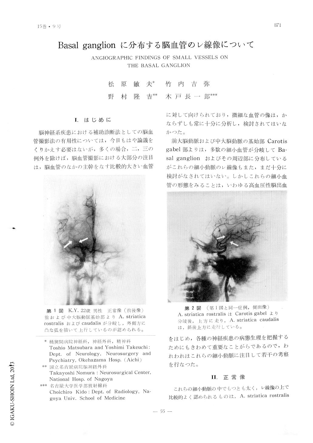
脳神経系疾患における補助診断法としての脳血管撮影法の有用性については,今日もはや論議をくりかえす必要はないが,多くの場合,二,三の例外を除けば,脳血管撮影における大部分の注目は,脳血管のなかの主幹をなす比較的大きい血管に対して向けられており,微細な血管の像は,かならずしも常に十分に分析し,検討されてはいなかつた。
前大脳動脈および中大脳動脈の基始部Carotis gabel部よりは,多数の細小血管が分岐してBa—sal ganglionおよびその周辺部に分布しているがこれらの細小動脈のレ線像もまた,まだ十分に検討がなされてはいない。しかしこれらの細小血管の形態をみることは,いわゆる高血圧性脳出血をはじめ,各種の神経疾患の病態生理を把握するためにもきわめて重要なことがらであるので,われわれはこれらの細小動脈に注目して若干の考察を行なつた。
Near the bifurcation of anterior and middle cerebral arteries (Carotid fork), numerous small arteries branch off which supply deep structures of the cerebral hemisphere. On ce-rebral angiograms, authors had taken special considerations on these arteries. The thicker artery among these vessels is A. striatica rostralis which bifurcate regressively from the supraoptic portion of anterior cerebral artery, and ascends with a laterally convexed curve on the A-P view of angiogram, and reaches on the anterior portion of corpus striatum. From middle cerebral artery, A striatica caudalis branches off also regressi-vely and ascends laterally and posteriorly to the A. striatica rostralis. Further, some arterial groups, Aa. pallido-thalamicae,pallido-striati-cae etc. bifurcate similarly from the middle cerebral artery, however, these vessels are too small to recognize separately on angiograms, authors had considered that it is clinically convenient to get together these small vascu-lar branches of middle cerebral artery with a group of A. striatica caudalis (A. striatica caudalis et al.).
In certain neurosurgical cases, including ce-rebral vascular disorders, some characteristic findings on A. striatica rostralis, caudalis et al. (mesial and lateral displacement, dissocia-tion of A. striatica rostralis and caudalis et al.) were obtained.

Copyright © 1963, Igaku-Shoin Ltd. All rights reserved.


