Japanese
English
- 有料閲覧
- Abstract 文献概要
- 1ページ目 Look Inside
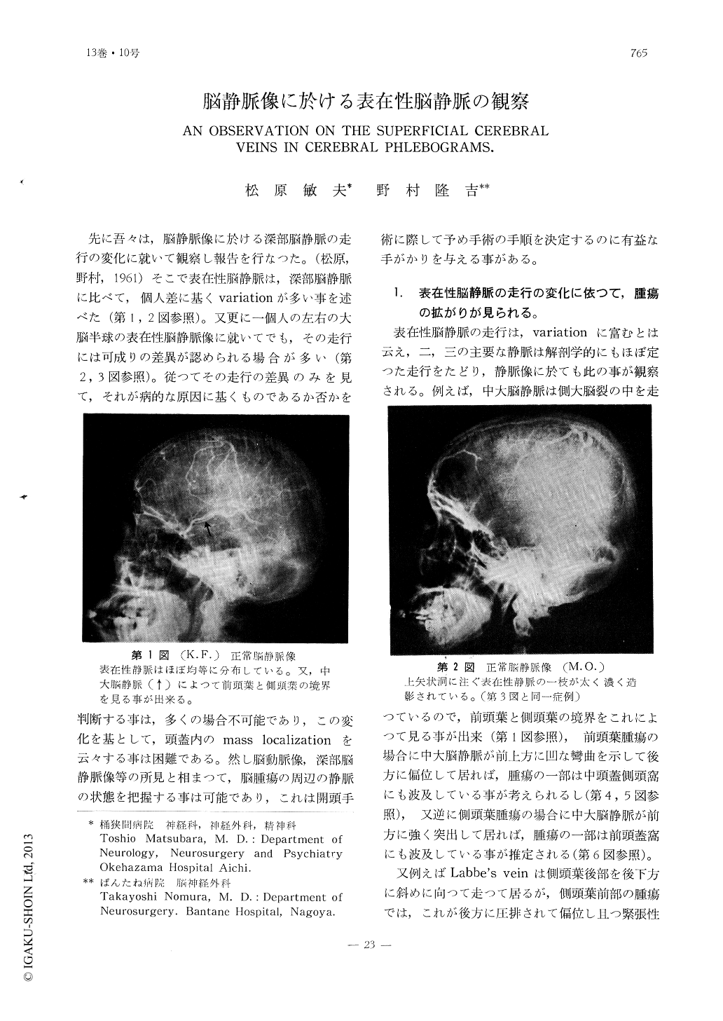
先に吾々は,脳静脈像に於ける深部脳静脈の走行の変化に就いて観察し報告を行なつた。(松原,野村,1961)そこで表在性脳静脈は,深部脳静脈に比べて,個人差に基くvariationが多い事を述べた(第1,2図参照)。又更に一個人の左右の大脳半球の表在性脳静脈像に就いてでも,その走行には可成りの差異が認められる場合が多い(第2,3図参照)。従つてその走行の差異のみを見て,それが病的な原因に基くものであるか否かを判断する事は,多くの場合不可能であり,この変化を基として,頭蓋内のmass localization を云々する事は困難である。然し脳動脈像,深部脳静脈像等の所見と相まつて,脳腫瘍の周辺の静脈の状態を把握する事は可能であり,これは開頭手術に際して予め手術の手順を決定するのに有益な手がかりを与える事がある。
(1) There are so many individual varia-tions in the coui se of superficial cerebral veins in each cerebral hemisphere that it seems impossible to distinguish the normal from the pathologic cerebral phlebograms. However, authors could have an observation about the characters of superficial veins surrounding the intracranial space taking lesions.
(2) It was recognized that the poor cont-rast filling of superficial cerebral veins in one area suggests the existence of brain ede-ma in the area.
(3) The direction of dominant superficial venous flow can be changed by the increased intracranial pressure. Although, equally dis-tributed superficial cerebral venous flow is most frequently observed,possible variations of the direction of dominant superficial venous flow are as follows ;
a) direction of dominant superficial vein to the sinus sagittalis superior, b) to sinus transversus, and c) to sinus caverno sus. The statistical observation on the direction of dominant superficial venous flow resulted that the b) and c) above mentioned have much relations to pathologic etiologies probably produced by the increased intracranial pres-sure.

Copyright © 1961, Igaku-Shoin Ltd. All rights reserved.


