Japanese
English
- 有料閲覧
PPV購入案内
1,320円 (1,200円+税10%) こちらは、1291ページから1297ページの文献です。購入には医書.jp本会員登録が必要です。
会員登録完了後に改めて上記「購入サイトへ」を選択してください。
または「購入サイトへ」選択後に「購入ログイン」、「新規会員登録」の順に進んでください。
会員登録について詳しくはをご確認ください。
- Abstract 文献概要
- 1ページ目 Look Inside
I.はじめに
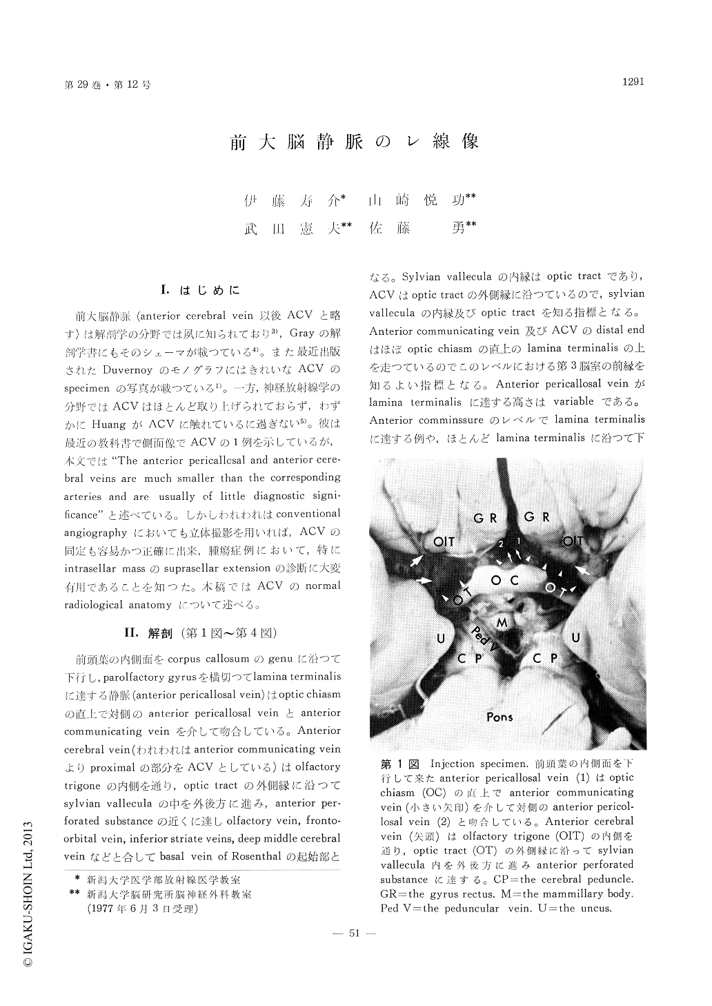
前大脳静脈(anterior cerebral vein以後ACVと略す)は解剖学の分野では夙に知られており3),Grayの解剖学書にもそのシェーマが載つている4)。また最近出版されたDuvernoyのモノグラフにはきれいなACVのspecimenの写真が載つている1)。一方,神経放射線学の分野ではACVはほとんど取り上げられておらず,わずかにHuangがACVに触れているに過き.ない5)。彼は最近の教科書で側面像でACVの1例を示しているが,本文では"The anterior pericallosal and anterior cere—bral veins are much smaller than the correspondingarteries and are usually of little diagnostic signi—ficance"と述べている。しかしわれわれはconventionalangiographyにおいても立体撮影を用いれば,ACVの同定も容易かつ正確に出来,腫瘍症例において,特にintrasellar massのsuprasellar extensionの診断に大変有用であることを知つた。本稿ではACVのnormalradiological anatomyにっいて述べる。
The anatomy of the anterior cerebral vein isdescribed with injection specimens. Radiological appearance of the anterior cerebral vein is illustrated with conventional angiograms. Stereoscopic films are indispensable to identify the anterior cerebral vein accurately. The anterior communicating vein or the distal end of the anterior cerebral vein indicates the anterior wall of the lower portion of the third ventricle. The anterior cerebral vein is very useful in defining the suprasellar extension of an intrasellar mass.
Copyright © 1977, Igaku-Shoin Ltd. All rights reserved.