Japanese
English
- 有料閲覧
- Abstract 文献概要
- 1ページ目 Look Inside
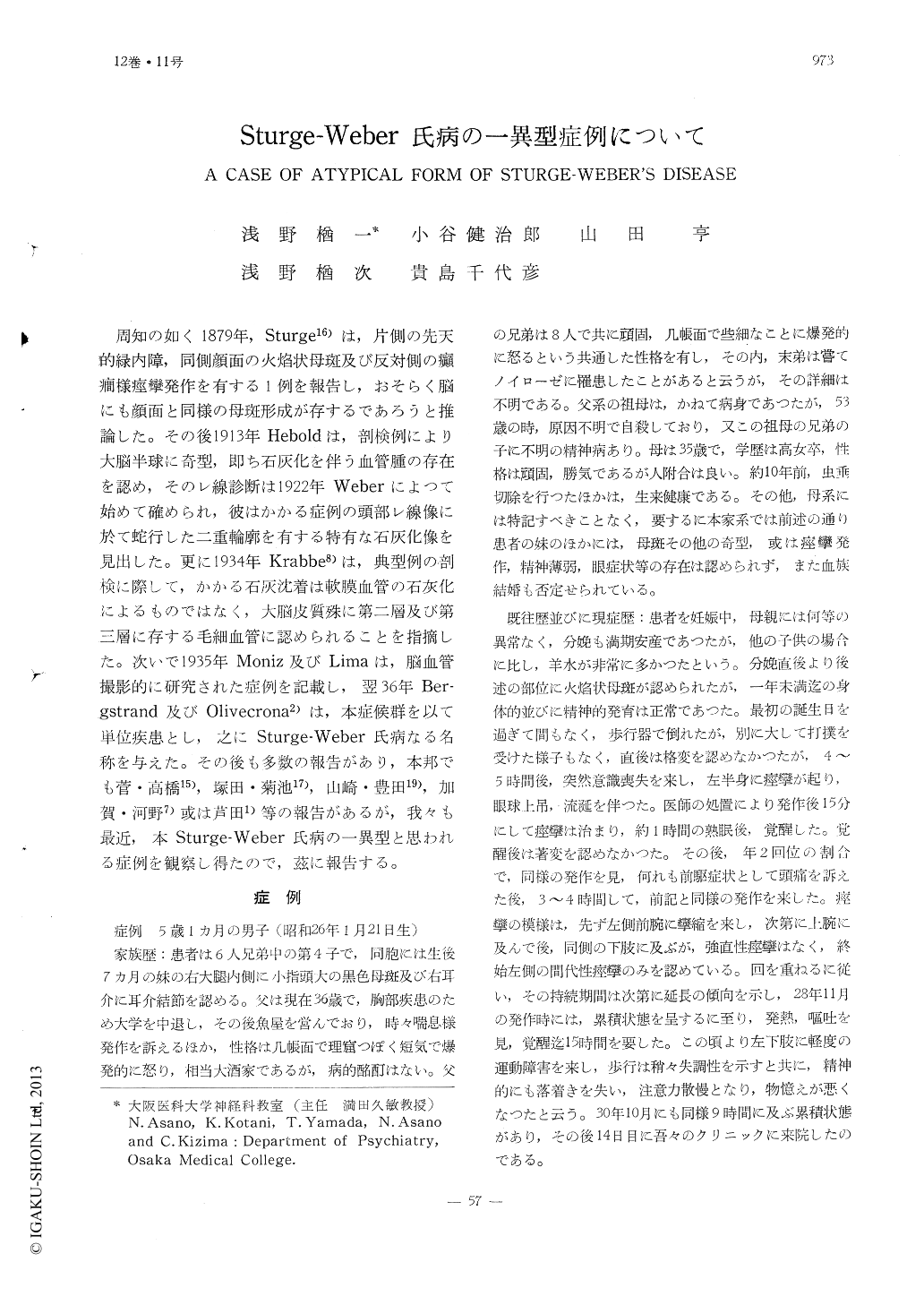
周知の如く1879年,Sturge16)は,片側の先天的緑内障,同側顔面の火焔状母斑及び反対側の癲癇様痙攣発作を有する1例を報告し,おそらく脳にも顔面と同様の母斑形成が存するであろうと推論した。その後1913年Heboldは,剖検例により大脳半球に奇型,即ち石灰化を伴う血管腫の存在を認め,そのレ線診断は1922年Weberによつて始めて確められ,彼はかかる症例の頭部レ線像に於て蛇行した二重輪廓を有する特有な石灰化像を見出した。更に1934年Krabbe8)は,典型例の剖検に際して,かかる石灰沈着は軟膜血管の石灰化によるものではなく,大脳皮質殊に第二層及び第三層に存する毛細血管に認められることを指摘した。次いで1935年Moniz及びLimaは,脳血管撮影的に研究された症例を記載し,翌36年Ber-gstrand及びOlivecrona2)は,本症候群を以て単位疾患とし,之にSturge-Weber氏病なる名称を与えた。その後も多数の報告があり,本邦でも菅・高橋15),塚田・菊池17),山崎・豊田19),加賀・河野7)或は芦田1)等の報告があるが,我々も最近,本Sturge-Weber氏病の一異型と思われる症例を観察し得たので,茲に報告する。
Case report was made on a 5 year-and-one month-old boy who was born with red naevus (Naevus vasculosus) on right-side of his face and in lower extremity of left side, and also, with blue naevus (Naevus caeruleus) in upper extremities bilaterally as well as in the back and abdomen. History revealed that he has been a subject of epileptic fits of Jacksonian type appearing approximately once a year over left side of body ever since one year of age. As he had gone through status epileptics at the age of 3, he gradually began to show restlessness and impairments of both atten-tion-span and memory-recording. He had I.Q. of 62 as determined by W.I.S.C.-I.Q.-test. Family history revealed neither consangui-nity nor presence of other types heredo-fami-liar factors except that the youngest sister had black naevus (benign melanoepithelioma) along right thigh, and right auricular tuber-cle. Patient has, in addition to the afore-mentioned symptoms, developed unilateral hypertrophy of left lower extremity. On the other hand, ophthalmic lesions including glau-coma have not been observed.

Copyright © 1960, Igaku-Shoin Ltd. All rights reserved.


