Japanese
English
- 有料閲覧
- Abstract 文献概要
- 1ページ目 Look Inside
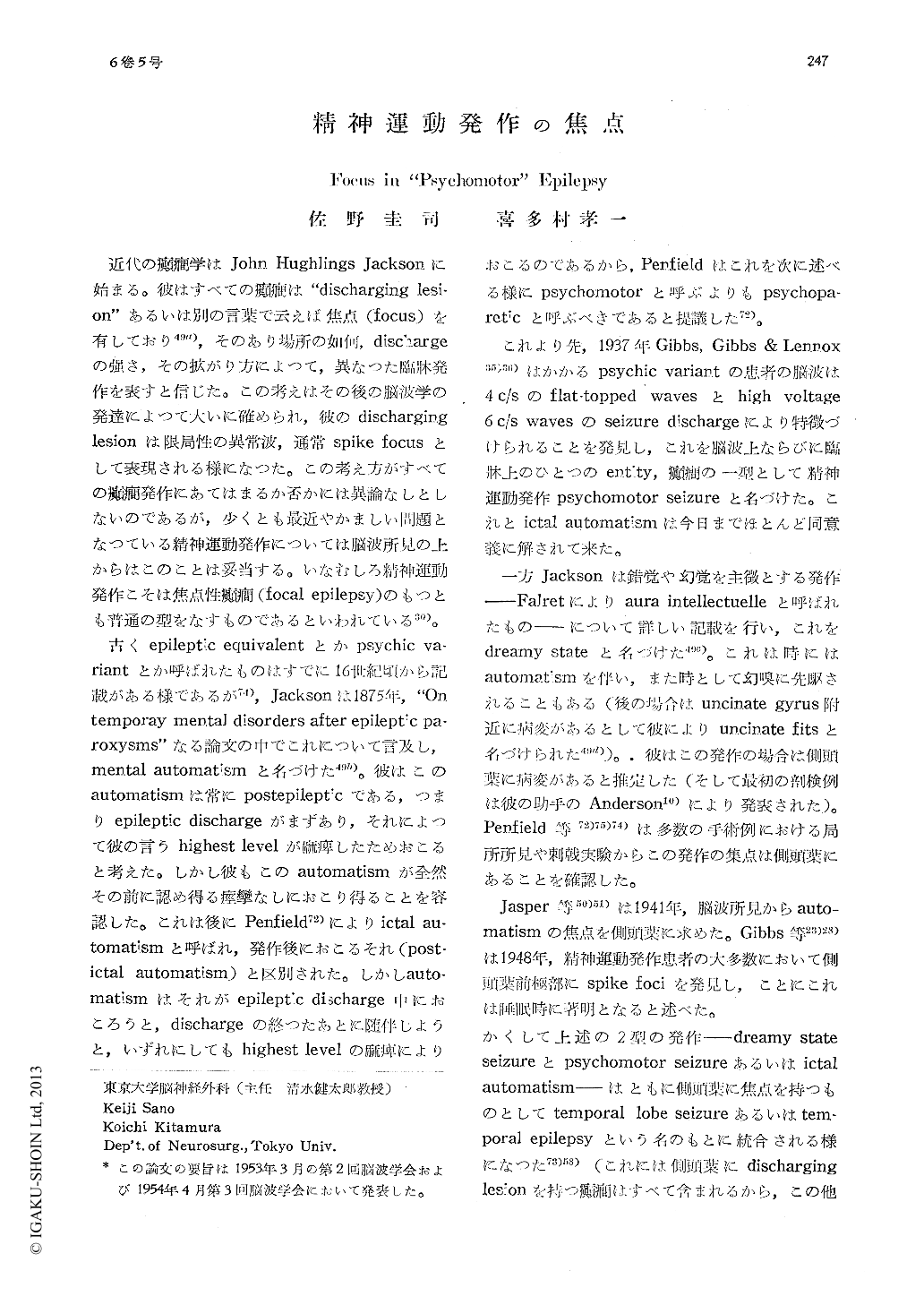
近代の癲癇学はJohn Hughlings Jacksonに始まる。彼はすべての癲癇は"discharging lesi-on"あるいは別の言葉で云えば焦点(focus)を有しており49a),そのあり場所の如何,dischargeの強さ,その拡がり方によつて,異なつた臨牀発作を表すと信じた。この考えはその後の脳波学の発達によつて大いに確められ,彼のdischarginglesionは限局性の異常波,通常spike focusとして表現される様になつた。この考え方がすべての癲癇発作にあてはまるか否かには異論なしとしないのであるが,少くとも最近やかましい問題となつている精神運動発作については脳波所見の上からはこのことは妥当する。いなむしろ精神運動発作こそは焦点性癲癇(focal epilepsy)のもつとも普通の型をなすものであるといわれている30)。
古くepileptic equivalentとかpsychic va-riantとか呼ばれたものはすでに16世紀頃から記載がある様であるが74),Jacksonは1875年,"Ontemporay mental disorders after epileptic pa-roxysms"なる論文の中でこれについて言及し,mental automatismと名づけた49b)。彼はこのautomatismは常にpostepilepticである,つまりepileptic dischargeがまずあり,それによつて彼の言うhighest levelが麻痺したためおこると考えた。しかし彼もこのautomatismが全然その前に認め得る痙攣なしにおこり得ることを容認した。これは後にPenfield72)によりictal au-tomatismと呼ばれ,発作後におこるそれ(post-ictal automatism)と区別された。しかしauto-matismはそれがepileptic discharge中におころうと,dischargeの終つたあとに随伴しようと,いずれにしてもhighest levelの麻痺によりおこるのであるから,Penfieldはこれを次に述べる様にpsychomotorと呼ぶよりもpsychopa-reticと呼ぶべきであると提議した72)。
(1) The authors reported on a new techni-que using long insulated needles inserted into the depth of the skull. This has proved to be particularly useful for detecting spike foci in "psychomotor" or temporal lobe epilepsy, when the needle tips were placed on the po-sterior edge of the base of the pterygoid pro-cess (medial infratemporal lead) and on the bony wall in the proximity of the anterior portion of the crista infratemporalis (anterior infratemporal lead).
(2) Of 44 cases with psychomotor seizures thus examined, marked spiking was noted in the medial infratemporal lead in 36 cases, in the anterior infratemporal lead in 5 cases, and in both medial and anterior leads withequal potential in 3 cases. 12 of these patients underwent craniotomy, 3 of them bilaterally.
(3) Spike foci were found in the subiculum (regiones prosubicularis & subicularis) in 8 occasions ; in the temporal cortex, mainly the polar area, in 4 occasions ; and in the amyg-daloid complex in one case.
(4) Microscopic examination of the hippo-campi excised in those cases with subiculum foci revealed so-called Ammon's horn sclerosis. Clinically the cases developed psychomotor seizures (not postictal) after the onset of the-ir habitual convulsive seizures (secondary psy-chomotor seizure). These findings suggest that in these cases sclerosis of the cornu Ammonis has been formed in the course of repeated convulsions and spike foci might de-velop around the sclerosed area, most likely in the subicular region, just as epileptogenic foci may develop around the cortical scar to cause posttraumatic epilepsy. These subiculum foci may bombard the diencephalon through the fornix on one hand and may send impulses to the temporal areas on the other.
(5) Besides these findings many wave pat-terns were noted in these areas. In the subicu-lum, 2-3c/s wave bursts, 12-15c/s positive spikes, 6-7c/s positive spikes, 4c/s flat-topped waves (during induced sleep), 6c/s wave bursts (during induced sleep) and random positive spikes were found. Random negative spikes and 4c/s flat-topped waves were also seen in the hippocampus proper.
(6) Strychnine neuronography was done dur-ing operation. Strychninization of the tempo-ral tip produced marked negative spikes there, which were conducted, after repetition of the firing, to the amygdaloid complex and the hi-ppocampus (and by far less markedly to the subiculum) as positive deflections. This was also the case with spontaneous negative spiking in the temporal pole. In the course of such repetitive spiking, the hippocampus and the amygdaloid complex were apparently activated and showed positive spiking of 2c/s rhythm, as if it were their proper rhythm. This aut-omatism of the electrical activity of these structures was suggestive of a possible bear-ing on emotional reactions to any given stimuli.
The hippocampal formation (the hippocam-pus, dentate fascia and the subiculum) did not show so-called strychine spikes on strych-ninization. But occasionally the hippocampus showed random large deflection, on inserting electrodes, which finally developed to 2c/s wave bursts. It was sometimes seen that stry-chninization of the hippocampal formation activated spont aneous spiking in the temporal cortex and the amygdaloid area without pro-duing appreciable spiking in themselves.

Copyright © 1954, Igaku-Shoin Ltd. All rights reserved.


