Japanese
English
- 有料閲覧
- Abstract 文献概要
- 1ページ目 Look Inside
I.緒言
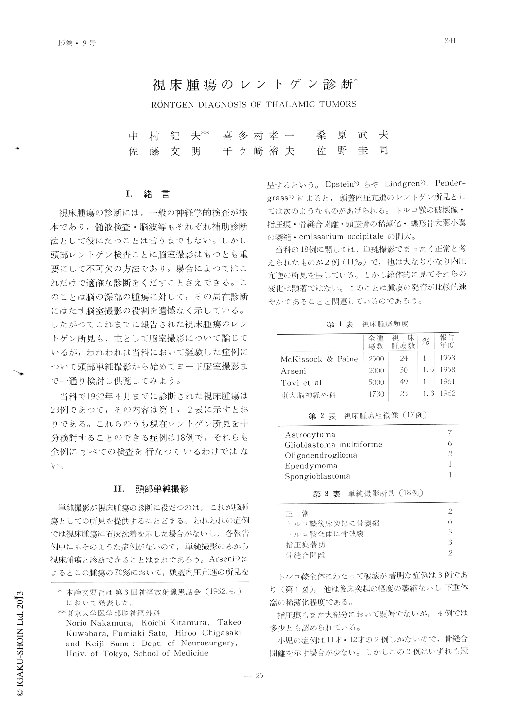
視床腫瘍の診断には,一般の神経学的検査が根本であり,髄液検査・脳波等もそれぞれ補助診断法として役にたつことは言うまでもない。しかし頭部レントゲン検査ことに脳室撮影はもつとも重要にして不可欠の方法であり,場合によつてはこれだけで適確な診断をくだすことさえできる。このことは脳の深部の腫瘍に対して,その局在診断にはたす脳室撮影の役割を遺憾なく示している。したがつてこれまでに報告された視床腫瘍のレントゲン所見も,主として脳室撮影について論じているが,われわれは当科において経験した症例について頭部単純撮影から始めてヨード脳室撮影まで一通り検討し供覧してみよう。
当科で1962年4月までに診断された視床腫瘍は23例であつて,その内容は第1, 2表に示すとおりである。これらのうち現在レントゲン所見を十分検討することのできる症例は18例で,それらも全例にすべての検査を行なつているわけではない。
Among 1730 cases of intracranial tumors in the neurosurgical department of the Univer-sity of Tokyo, 23 were diagnosed as the tumor of the thalamus from the results of X-ray examination, the clinical examination or biopsy and antopsy.
The authors demonstrated, summarized and discussed all of the X-ray films of these tumors.
Many of the plain craniograms showed less dominant destruction of the skull which might be attributed to the rapid developement of the tumor.
In cerebral angiograms, carotid phlebograms were. most valuable for diagnosis of the tu-mor, but interpretation of the findings needed much experience and skill.
The widening of the so-called venous angle or the abnormal vascularization at the branch of the thalamostriate vein was the most pa-thognomonic sign.
All of the abnormal vascularization was revealed to be due to malignant glioma in autopsy.
In 14 cases of pneumoventriculograms, 7 sho-wed shadow of the tumor in the lateral ven-tricle, which occupied the central part or the trigone. The central part of the ipsilateral ventricle was elevated and flattened, and smaller than that of the contralateral one.

Copyright © 1963, Igaku-Shoin Ltd. All rights reserved.


