Japanese
English
- 有料閲覧
- Abstract 文献概要
- 1ページ目 Look Inside
I.はじめに
てんかんについての電気現象は従来より色々と研究されて来た。しかし直流電位の面から分析した研究は決して多いものではない。それでも発作波に伴う直流電位の研究はGoldringら(1950)4)以来綿々と続けられて来た。ところが,てんかん焦点と直流電位の関係を調べたものとなると,ずつと数が少ない。しかもそのほとんどは,直流電位成分のうちの100μVオーダーの変動成分である緩変動電位(slowly changing Potential. SCP)についての研究である15)。直流電位のもう一つの重要な成分,定常電位(stationary potential.以下SPと略す)──この電位は神経細胞の膜電位に起源する+10〜20mVの,通常の状態ではきわめて定常な電位であり,器質的脳障害により増減する9)11)12)──とてんかん焦点との関係をみた研究はほんの数篇でしかない8)10)13)。
本論文の目的は,従来ないがしろにされて来たてんかん焦点におけるSPの変動とその病態生理学的意義を明らかにすることである。そのために次のような検討を行なつた。すなわち臨床的には,①グリオーマに比べ病態的に単純である髄膜腫について,そのSP分布異常がてんかん発生率に関係しうるものかどうか,②焦点性てんかんにSP分布異常がみられるか,みられるとすればその頻度はどの程度か,③焦点性てんかんの場合脳表にもSP分布異常が証明できるか,を検討した。
The electrical activities of the brain recorded with macroelectrodes can be classified into 3 main categories:
(1) What is called EEG, (2) the slowly changing potential (SCP) which is of the oder of microvolts (ranging from 50μV to several millivolts) and is slower than 0.5 Hz, (3) and the stationary potential which is of the oder of millivolts and is very stable as the name implies. The last two types of potentials are often called DC potentials or steady potentials.
The stationary potential requires a special record-ing method which consists of calomel half-cell electrodes, salt bridges and a high impedance (30,000 Megohms) DC amplifier.
Recently this system is connected with an auto-matic scanner which detects each voltage difference of 5mV.
So far we have measured the SP in 294 cases. Normal controls show a symmetrical voltage dis-tribution with the highest potential in the vertex, the range being 10-20 mV when the reference elect-rode is on the nasion. If an organic lesion is located in the cerebral hemisphere, the SP shows various changes according to the location of the lesion.
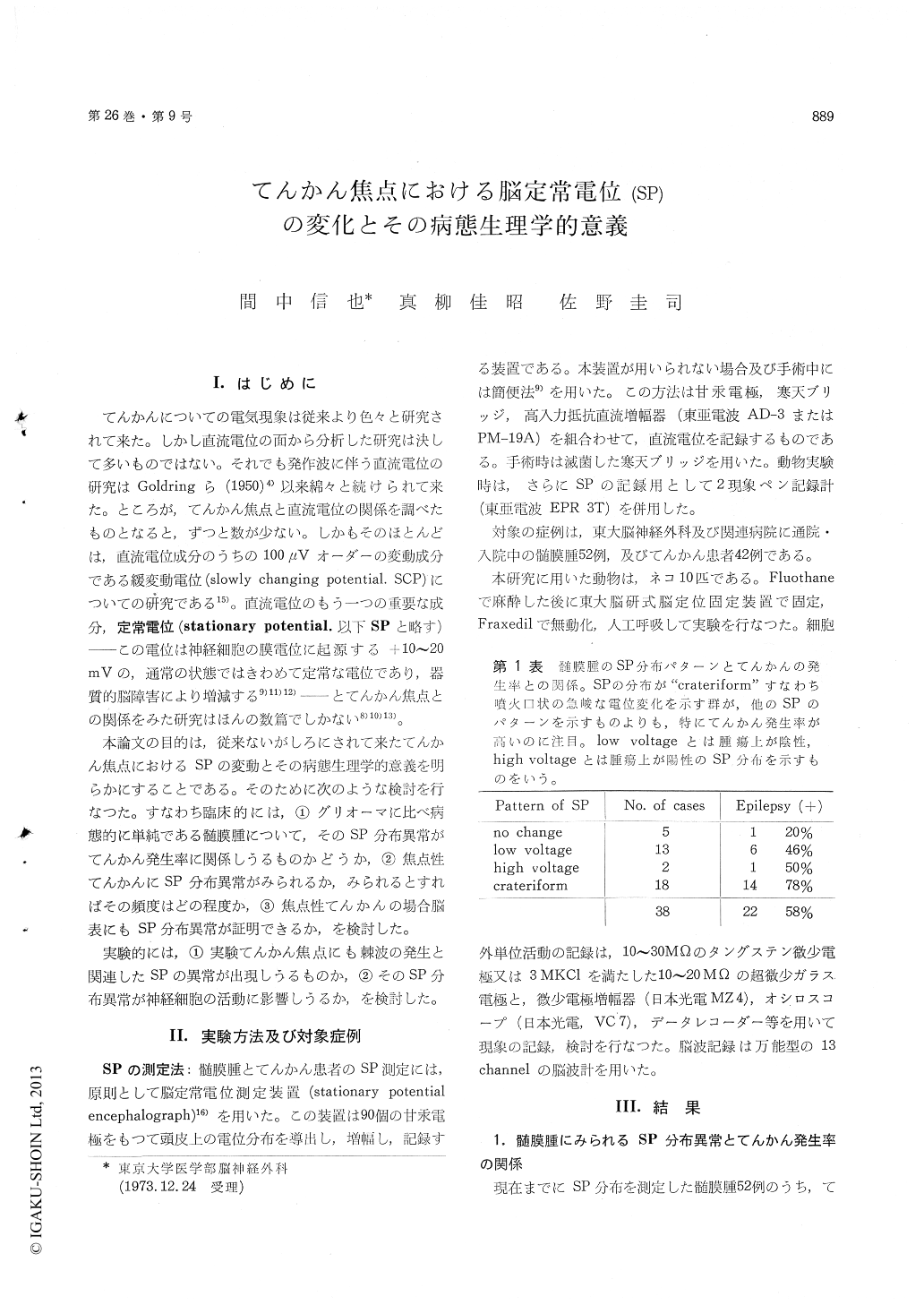
Cases of meningioma show of ten (about 50%) what we call a "crateriform potential change" which consists of negativity in the center (over the tumor) and positivity surrounding it (over the adjacent cortex).
78 per cent of those cases showing the crateri-form potential change had epileptic attacks, focal or generalized, whereas cases showing negativity over the tumor without surrounding positivity had seizu-res in 46 per cent and cases without SP changes had seizures only in 20 per cent of the cases (Table 1). This may mean that the steep SP potential gradient is irritable to the cortex, working in the source and sink relationship.
Cases of cryptofocal or non-focal epilepsy usually show a normal SP distribution, whereas cases of focal epilepsy often exhibit SP changes (Table 2 and 3). About 53 per cent of the latter show low (negative) SP on the foci. This suggest the ex-istence of an organic lesion involving the cortex.
The cortical scar of one of the cases of traumatic epilepsy shows negativity of about 15 mV as com-pared with the surrounding normal cortex. EEGspiking is seen in the surrounding cortex. The cortex of one of the cases of ectopic gray matter shows negativity of about 4 mV as compared with the surrounding area (Figure 1 and 2).
The experimental epileptogenic focus produced by cortical application of penicillin shows a steep stationary potential (SP) gradient with negativity in the applied area and positivity in the surrounding cortex which exhibits spiking (Figure 3). This is in accordance with the report of Mahnke and Ward.
Artificial SP change by applied cortical polari-zation in cats exerted a remarkable influence on the frequency of neuronal uhit discharges (Figure 4).
Conclusion :
1) SP change on the focus produces hyperexi-tability, working in source and sink relationship, and facilitates production of sezure discharge (Figure 5).
2) Measuring the SP change on the scalp and/or on the cortex is useful for diagnosis and operative treatment of the focal epilepsy.

Copyright © 1974, Igaku-Shoin Ltd. All rights reserved.


