Japanese
English
- 有料閲覧
- Abstract 文献概要
- 1ページ目 Look Inside
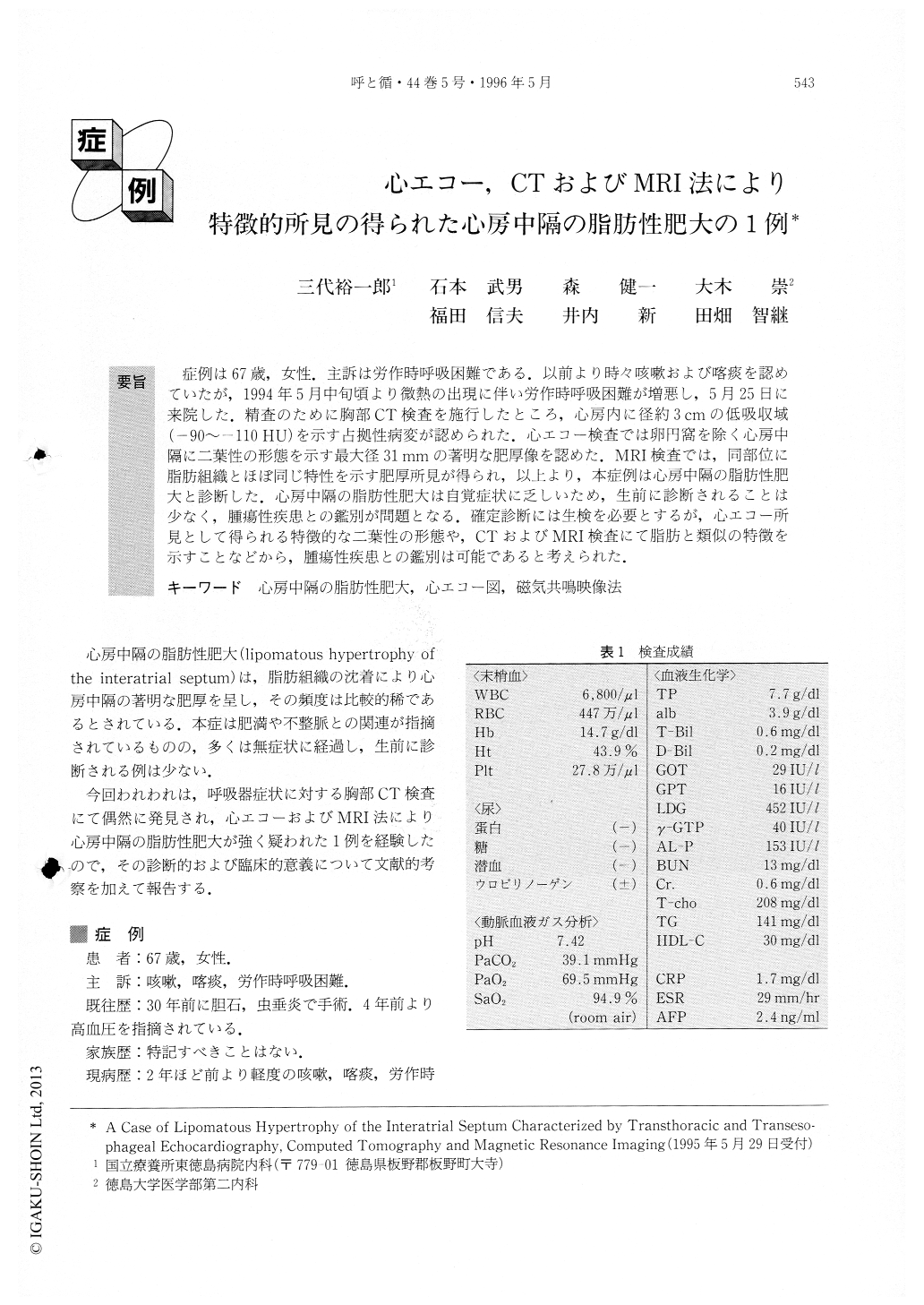
症例は67歳,女性.主訴は労作時呼吸困難である.以前より時々咳嗽および喀痰を認めていたが,1994年5月中旬頃より微熱の出現に伴い労作時呼吸困難が増悪し,5月25日に来院した.精査のために胸部CT検査を施行したところ,心房内に径約3cmの低吸収域(−90〜−110HU)を示す占拠性病変が認められた.心エコー検査では卵円窩を除く心房中隔に二葉性の形態を示す最大径31mmの著明な肥厚像を認めた.MRI検査では,同部位に脂肪組織とほぼ同じ特性を示す肥厚所見が得られ,以上より,本症例は心房中隔の脂肪性肥大と診断した.心房中隔の脂肪性肥大は自覚症状に乏しいため,生前に診断されることは少なく,腫瘍性疾患との鑑別が問題となる.確定診断には生検を必要とするが,心エコー所見として得られる特徴的な二葉性の形態や,CTおよびMRI検査にて脂肪と類似の特徴を示すことなどから,腫瘍性疾患との鑑別は可能であると考えられた.
We described a case of lipomatous hypertrophy of the interatrial septum characterized by transthoracic and transesophageal echocardiography, computed tomogra-phy (CT) and magnetic resonance imaging (MRI). A 67-year-old woman who complained of cough, sputum and dyspnea on effort visited our hospital. Her chest CT showed a low density mass with 3cm diameter (-90 to-110 Hounsfield units) at the level of the atrial septum. In order to differentiate the mass from myxoma, thrombus and other tumors, transthoracic andtransesophageal echocardiography and MRI were per-formed. Echocardiography identified the characteristic bilobed appearance of the atrial septum with relative sparing of the fossa ovalis, but right and left ventricular inflow obstruction to blood flow wasn't proved hemodvnamically. MRI identified the adipose nature of the tissue on both T1- and T2-weighted images, and appeared contiguous with the normal epicardial fat. The diagnosis of lipomatous hypertrophy of the inter-atrial septum may be inferred from the density of the mass determined by either CT or MRI, and the morphol-ogy of the characteristic bilobed appearance by echocardiography.

Copyright © 1996, Igaku-Shoin Ltd. All rights reserved.


