Japanese
English
- 有料閲覧
- Abstract 文献概要
- 1ページ目 Look Inside
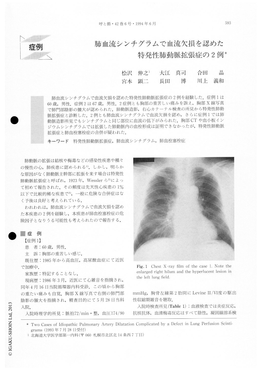
肺血流シンチグラムで血流欠損を認めた特発性肺動脈拡張症の2例を経験した.症例1は60歳,男性.症例2は67歳,男性.2症例とも胸部の重苦しい痛みを訴え,胸部X線写真で肺門部陰影の腫大が認められた.肺動脈造影,右心カテーテル検査の所見から特発性肺動脈拡張症と診断した.2例とも肺血流シンチグラムで血流欠損を認め,さらに症例1では肺動脈造影所見でもシンチグラムと同じ部位に血流の低下がみられた.胸部CTや血小板インジウムシンチグラムでは拡張した肺動脈内の血栓形成は証明できなかったが,特発性肺動脈拡張症と肺血栓塞栓症の合併が疑われた.
A 60-year-old male and a 67-year-old male were admitted because of hilar enlargement seen on chest radiography. Both patients complained of chest oppres-sion and were diagnosed as IAPD on the basis of the findings of the pulmonary angiogram and right heart catheterization. Moreover pulmonary angiogram of the latter case demonstrated decreased perfusion in the right upper lung field. Although chest computed tomo-graphy and lung scintigraphy by radiolabelled platelet could not detect the formation of thrombus in the enlarged main pulmonary artery, both cases were suspected to have pulmonary embolism secondary to IPAD.

Copyright © 1994, Igaku-Shoin Ltd. All rights reserved.


