Japanese
English
- 有料閲覧
- Abstract 文献概要
- 1ページ目 Look Inside
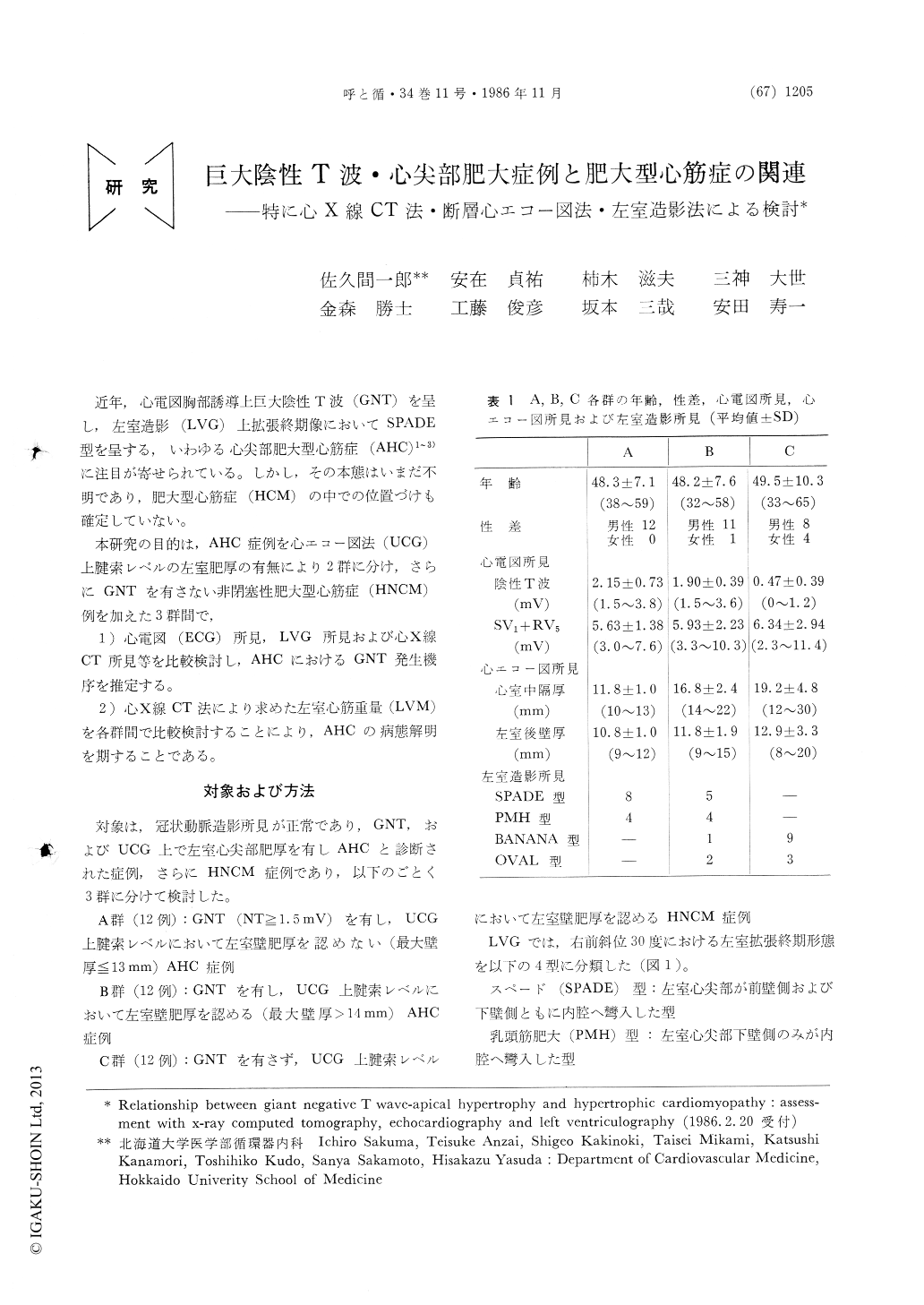
近年,心電図胸部誘導上巨大陰性T波(GNT)を呈し,左室造影(LVG)上拡張終期像においてSPADE型を呈する,いわゆる心尖部肥大型心筋症(AHC)1〜3)に注目が寄せられている。しかし,その本態はいまだ不明であり,肥大型心筋症(HCM)の中での位置づけも確定していない。
本研究の目的は,AHC症例を心エコー図法(UCG)上腱索レベルの左室肥厚の有無により2群に分け,さらにGNTを有さない非閉塞性肥大型心筋症(HNCM)例を加えた3群間で, 1)心電図(ECG)所見,LVG所見および心X線CT所見等を比較検討し,AHCにおけるGNT発生機序を推定する。 2)心X線CT法により求めた左室心筋重量(LVM)を各群間で比較検討することにより,AHCの病態解明を期することである。
To assess a relationship between giant negative T wave (GNT) and left ventricular (LV) hypertrophic patterns, and to clarify the pathogenesis of apical hypertrophic cardiomyopathy (AHC), 12 patients (pts) with AHC (group A: GNT (+), mid-ventricular hypertrophy at chorda level in echocardiography (mVH) (-)) and 24 pts with non-obstructive hyper-trophic cardiomyopathy (group B: GNT (+), mVH (+), n=12, and group C: GNT (-), mVH (+), n=12) were studied with multi-slice ECG gated X-ray computed tomography (MSECT) and left ventriculogra-phy (LVG).
In MSECT images pts in group A showed LV hyper-trophy (LVH) localized at the apex and pts in group B showed LVH relatively thicker at the apex ; however pts in group C had more severe LVH spreading towards the base. End-diastolic LVG patterns were spade or spade-like pattern in group A, although non-spade pat-tern in group C. Left ventricular mass (LVM) cal-culated from MSECT was significantly heavier in group C than in groups A and B. In groups A and B there was a wide variation in LVM from as light as that in normal controls to as heavy as that in group C.
In conclusion, LVH relatively localized at the apex was assumed to generate GNT. AHC pts showed a wide variation in the severity of LVH and their patho-geneses might be different.
Copyright © 1986, Igaku-Shoin Ltd. All rights reserved.