Japanese
English
- 有料閲覧
- Abstract 文献概要
- 1ページ目 Look Inside
近年,核医学領域の進歩は著しく,循環器疾患の分野においてもこの進歩は例外でない。とくに,SinglePhoton Emission Computed Tomography (SPECT)の開発により三次元画像の作製が可能となり,心筋イメージングの臨床応用ではすでに高い評価を得るに至っている1〜5)。心プールイメージングの分野でもMultigate法6)や位相解析法Phase Analysis7)の開発により,各種心機能指標を用いた心疾患の病態解析の試みが数多く報告されている8〜11)。しかし,SPECTのこの領域への応用はいまだ僅少にとどまる。
著者らは,RI心プール法にSPECTを応用することで心臓を三次元的にとらえ,また位相解析法を行うことで,空間的かつ時間的な面からの局所心機能評価を試みた。本稿では,心筋梗塞例および特発性心筋症例を対象とした本法の有用性について述べる。
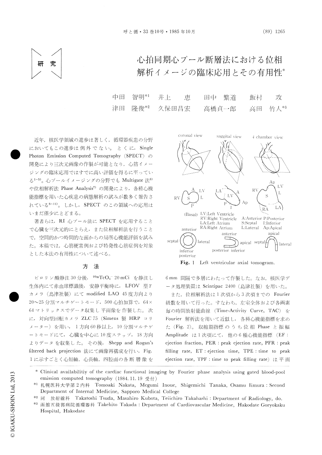
Phase analysis in ECG gated blood-pool angio-grams has been recently employed in clinical study for evaluating global and regional cardiac function. However, equilibrium radionuclide angiograms collected in one dimensional projection have a limi-tation to detect localization of the abnormality pre-cisely. In order to resolve this problem, the pool images were reconstructed three-dimensionally with "coronal", "sagittal", and "four chamber" tomo-grams by using gated blood-pool emission computed tomography (GPECT). In addition, fundamentaland higher order Fourier analyses were applied to every image to assess the spatial and temporal move-ment of the regional walls. In this paper, we de-scribed two typical cases and the availability of this method.
Case 1 is a 56 year-old man in whom myocar-dial infarction of the anterior, lateral, apical and posterior walls was diagnosed with the ECG, TI-201 myocardial tomograms, coronary angiography and contrast ventriculography. Although the con-ventional planar images could show low amplitude and phase delay at the lateral site of the left ventricule, a more detailed estimation of the local function could not be done because of an ovarlapped blood pool due to the two-dimensional display. In contrast, the functional images by GPECT were able to express phase delay and low amplitude at the anterior, lateral, apical and poste-rior portions in the coronal, sagittal and four cham-ber sections, which coincided with their infarc-tion areas, respectively.
Case 2 is a 57 year-old man with hypertrophic obstructive cardiomyopathy, diagnosed with the echocardiogram and contrast ventriculography. Higher order functional images in the LAO view suspected systolic hyperfunction and diastolic dysfunc-tion. Applying GPECT to this case, a more severe degree of damage in diastole was observed, especial-ly in the left ventricular outflow tract, from the functional images of PFR (peak filling rate) and TPF (time to peak filling rate) in the sagittal and four chamber sections. In conclusion, it was suggested that the cardiac functional images reconstructed three-dimensionally by GPECT might be useful for a more detailed and more precise evaluation of the regional wall motion in various cardiac disorders, overcoming anatomical demerits in the conventional planar image.
Copyright © 1985, Igaku-Shoin Ltd. All rights reserved.