Japanese
English
- 有料閲覧
- Abstract 文献概要
- 1ページ目 Look Inside
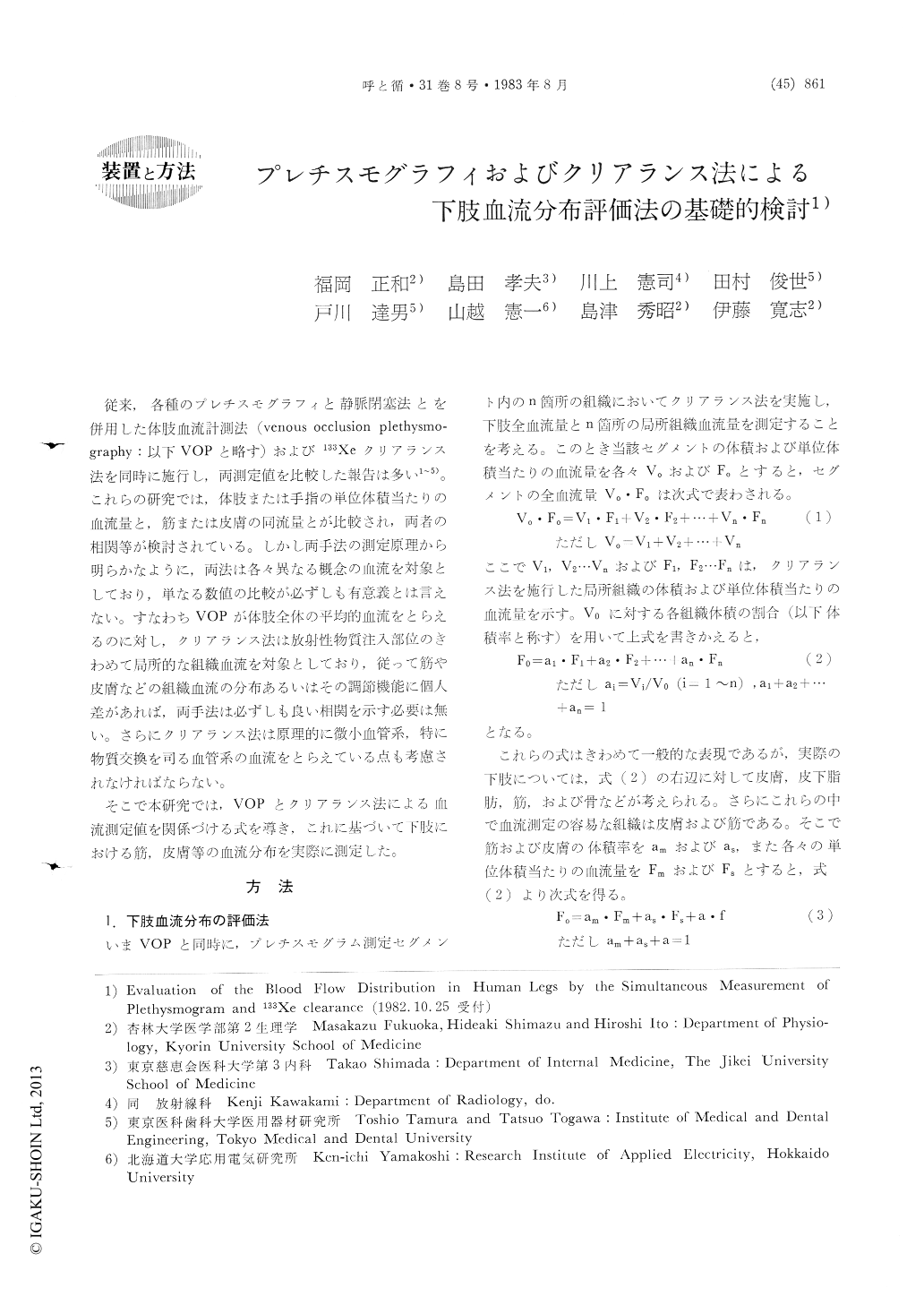
従来,各種のプレチスモグラフィと静脈閉塞法とを併用した体肢血流計測法(venous occlusion plethysmo—graphy:以下VOPと略す)および133Xeクリアランス法を同時に施行し,両測定値を比較した報告は多い1〜5)。これらの研究では,体肢または手指の単位体積当たりの血流量と,筋または皮膚の同流量とが比較され,両者の相関等が検討されている。しかし両手法の測定原理から明らかなように,両法は各々異なる概念の血流を対象としており,単なる数値の比較が必ず唱も有意義とは言えない。すなわちVOPが体肢全体の平均的血流をとらえるのに対し,クリアランス法は放射性物質注入部位のきわめて局所的な組織血流を対象としており,従って筋や皮膚などの組織血流の分布あるいはその調節機能に個人差があれば,両手法は必ずしも良い相関を示す必要は無い。さらにクリアランス法は原理的に微小血管系,特に物質交換を司る血管系の血流をとらえている点も考慮されなければならない。
そこで本研究では,VOPとクリアランス法による血流測定値を関係づける式を導き,これに基づいて下肢における筋,皮膚等の血流分布を実際に測定した。
Blood flow distribution in human legs was evaluated by the simultaneous application of electrical admittance plethysmography and 133Xe clearances technique. The leg segment was considered to be composed of three parts ; the muscle, the skin, and the remaining tissue. The blood flows of the skin and the muscle (Fs and Fm) were measured by the clearance technique, while the mean total flow of the leg segment (Fo) was determined by the admittance plethysmography. Since these blood flows were expressed as the values per unit tissue volume, the following equation can be used to evaluate the blood flow distribution in the leg ; Fo=am・Fm+as・Fs+a・f, where am, as, and a were the volume ratios of the muscle, skin and the remaining tissue to the total leg segment, respectively. Thus, the blood flow distribution can be determined by the blood flow rates of the muscle (am・Fm/Fo), the skin (as・Fs/Fo), and the remaining tissues (a・f/Fo). From anatomical and X-ray findings, we calculated the mean values of the volume ratios ; am=0.65, as,=0.1 and a=0.25. Measurements were carried out on the legs of 17 healthy subjects (male ; 26-38 yr.) at rest in a supine position. The measured values (mean ±SD ; ml/min/100ml) of Fo, Fm and Fs were 2.22±0.77, 1.76±.0.72, and 10.0±5.7, respectively. Thus, the blood flow rates were 51, 41 and 8% in the muscle, in the skin and in the remaining tissue, respectively. The accuracy of this method and the significance of the blood flow in the remaining tissue (a・f) were discussed in detail.

Copyright © 1983, Igaku-Shoin Ltd. All rights reserved.


