Japanese
English
- 有料閲覧
- Abstract 文献概要
- 1ページ目 Look Inside
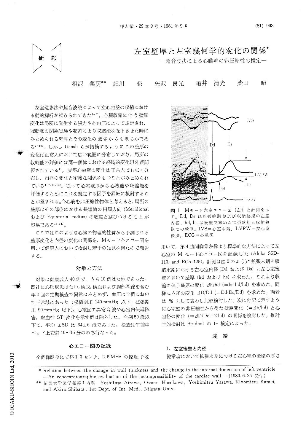
左室造影法や超音波法によって左心室壁の収縮における動的解析が試みられてきた1〜6)。心臓収縮に伴う壁厚変化は局所に発生する張力や心内圧によって規定され,冠動脈の閉塞実験や薬剤により収縮能を低下させた時にみとあられる壁厚とその変化の減少からも明らかである7〜10)。しかしGaashらが指摘するようにこの壁厚の変化は正常人において広い範囲に分布しており,局所の収縮能の評価には同一個体における経時的変化以外疑問視されている1)。実際心室壁の変化は正常人でも広く分布し,内径の変化と密接な関係をもつことがみとめられている4〜7,11,12)。従って心室壁厚から心機能や収縮能を評価するためにこれを規定する因子を詳細に検討することが望まれる。今心筋を非圧縮性物体と考えると,局所の壁厚はその部位における長短軸の円周方向(MeridionalおよびEquatorial radius)の収縮と結びつけることが容易である13,14)。
ここではこのような心臓の物理的性質から予測される壁厚変化と内径の変化の関係を,Mモード心エコー図を用いて健康人において検討し若干の知見を得たので報告する。
Assuming the cardiac wall as incompressible material, the change in the wall thickness during contraction can be related to the change in the left ventricular dimension. The pridicted relation was assessed in M-mode echocardiographic re-cordings in man (N=40).
The fractional change in wall thickness was found to be widely distributed. Such a wide distribution was also found in the fractional change of left ventricular dimension. The frac-tional change in wall thickness was found to be highly correlated with that of dimensional change as predicted from the property of the incompres-sibility (r=0.85, p<0.001). This result suggests that the incompressibility of cardiac wall seems to be proved by echocardiography. Abnormal regional contraction;hyper or hypokinesia, may be detected as a deviation from the regression equation obtained for the fractional change of wall thickness and the dimensional change.

Copyright © 1981, Igaku-Shoin Ltd. All rights reserved.


