Japanese
English
- 有料閲覧
- Abstract 文献概要
- 1ページ目 Look Inside
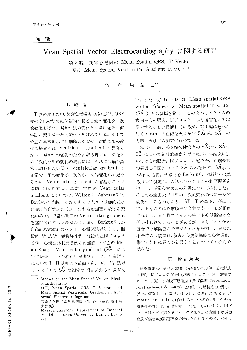
I.緒言
T波の変化の中,興奮伝播過程の変化即ちQRS波の変化のために付随的に起るT波の変化を二次的変化と呼び,QRS波の変化とは別に起るT波単独の変化は一次的変化と呼ばれている。そして心筋の異常を示す心筋傷害などの一次的なTの変化の場合にはVentricular gradientは異常となり,QRSの変化のために起る脚ブロックなどの二次的なTの変化の場合には,それに心筋の異常が加わらない限りVentricular gradientま正常で,Tの変化が一次的か二次的変化かを定めるのにVentricular gradientの有益なことが指摘されて来た。異常心電図のVentriculargradientについては,Wilson1),Ashman2),3),Bayley4)以来,かなり多くの人々の基礎的並びに臨床約研究があるが,何れも前額面に於ける変化のみで,異常心電図のVentricular gradientを空間的に扱つた者はなく,最近Berkun5)らがCube systemのベクトル心電図誘導法より,間歇的W.P.W.症候群4例,間歇的左脚ブロック6例,心室期外収縮8例の前額面,水平面のMe—an Spatial Ventricular gradient (SĜ)について報告し,また柏村6)が脚ブロック,心室肥大についてI, II誘導より前額面を,V2, V6誘導より水平面のSĜの測定の報告があるに過ぎない。
1) The magnitudes and directions of SÂQRS, SÂT and SĜ, and the relations among these three vectors were studied in abnormal electrocardiograms with ventricular hypertrophy, bundle branch block, subendocardial ischemia and injury, and myocardial infarction by the methods described in the first and second recports.
2) In left ventricular hypertrophy and left bundle block, SÂQRS was mostly directed to the left and posteriorly, and SÂT was mostly directed to the right, inferiorly and anteriorly. In right ventricular hypertrophy and right bundle branch block, SÂQRS was mostly directed to the right, inferiorly and anteriorly, and SÂT mostly to the left and posteriorly. In subendocardial ischemia and injury SÂQRS was mostly directed to the left, inferiorly and posteriorly, and SÂT mostly to the right, inferiorly and posteriorly. In myocardial infarction SÂQRS was mostly to the left and post-eriorly, but the direction tended to vary according to the location of infarct; especially SÂT was strongly affected by the location and phase of infarction. Furthermore, in most instances of left ventricular hypertrophy and left bundle branch block SÂT lay in the clockwise direction of SÂQRS in the frontal and horizontal planes, while in right ventricular hypertrophy and right bundle branch block approximately the reverse relations were observed.
3) Almost all cases of ventricular hypertrophy, bundle branch block and myocardial ischemia showed an increase in ∠(SÂQRS・ SÂT).
4) Ventricular hypertrophy and left bundle branch block showed an increase in ∠(SÂT・ SĜ). Some of right bundle branch block and myocardial infarction, however, showed an increase in ∠(SÂQRS・) SĜ) and some showed an increase in ∠(SÂT・SĜ) but subepicardial ischemia showed an increase in ∠(SÂQRS・SĜ). Moreover, ∠(SÂQRS・SÂT), ∠(SÂQRS・SĜ) and ∠(SÂT・SĜ) varied according to the directions and also to the magnitudes of SÂQRS, SÂT and SĜ in each dissease. Therefore, it is ina-dequate to evaluate SÂQRS, SÂT and SĜ themselves without considerlation of their correlations.
5) The magnitude of SĜ was generally decreased in the presence of myocardial ischemia. Howe-ver, in ventricular hypertrophy and bundle branch block, even they did not combine the myocardial ischemia, the magnitude of SĜ was decreased by the decrease in the magnitude of SÂQRS and SÂT, and the increase in the spatial angle.
6) In myocardial infarction SÂQRS, SÂT, SĜ and their spatial angles changed according to the location and phase of infarction. The change of SĜ was rather in parallel with SÂT, but the SÂT change was more remarkable than SĜ.
7) It was often observed that the direction of SĜ was normal in the frontal plane but abnormal in the horizontal, and vice versa. Therefore, it is unsatisfactory to observe the direction of SĜ only in the frontal plane. Abnormal directions of SĜ were found in 11 of 20 cases with ventricular hyp-ertrophy, in 13 of 20 cases with bundle branch block, 1 of 20 cases with subendocardial ischemia and injury, and 14 of 20 cases of myocardial infarction. However, all of the 3 cases of ventricular hyper-trophy with coronary (coved) T, 4 cases of bundle branch block with myocardial infarction, and 3 cases of subepicardial ischemia showed the abnormal direction of SĜ. These results indicate that if primary T wave changes occur as a result of disease of myocadium, the gradient will be abnormal.
8) Further, the differences between subendocardial and subepicardial ischemia were discussed and the clinical significance of SĜ was described in this report.

Copyright © 1958, Igaku-Shoin Ltd. All rights reserved.


