Japanese
English
- 有料閲覧
- Abstract 文献概要
- 1ページ目 Look Inside
- 参考文献 Reference
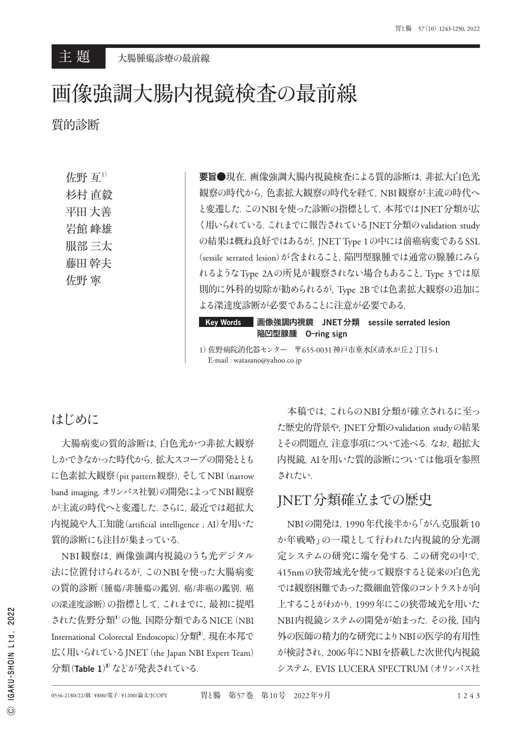
要旨●現在,画像強調大腸内視鏡検査による質的診断は,非拡大白色光観察の時代から,色素拡大観察の時代を経て,NBI観察が主流の時代へと変遷した.このNBIを使った診断の指標として,本邦ではJNET分類が広く用いられている.これまでに報告されているJNET分類のvalidation studyの結果は概ね良好ではあるが,JNET Type 1の中には前癌病変であるSSL(sessile serrated lesion)が含まれること,陥凹型腺腫では通常の腺腫にみられるようなType 2Aの所見が観察されない場合もあること,Type 3では原則的に外科的切除が勧められるが,Type 2Bでは色素拡大観察の追加による深達度診断が必要であることに注意が必要である.
Recently, the qualitative and quantitative diagnostic methods using image-enhanced colonoscopy have transitioned from the usage of non-magnified white light to magnified chromoendoscopic visualization and finally to NBI(narrow band imaging)visualization, which is the most commonly used method now. JNET(the Japan NBI Expert Team)classification is widely used in Japan as an index for diagnosis using magnified NBI. Although several validation studies for the JNET classification reported to date are generally favorable, it is essential to consider the following three points:1) JNET Type 1 lesions include pre-cancerous sessile serrated lesions, 2) Type 2A findings as observed in conventional adenomas may not be observed in depressed adenomas, and 3) surgical excision is recommended for Type 3 lesions, while Type 2B lesions require an in-depth diagnosis by additional magnified chromoendoscopic visualization.

Copyright © 2022, Igaku-Shoin Ltd. All rights reserved.


