Japanese
English
- 有料閲覧
- Abstract 文献概要
- 1ページ目 Look Inside
- 参考文献 Reference
- サイト内被引用 Cited by
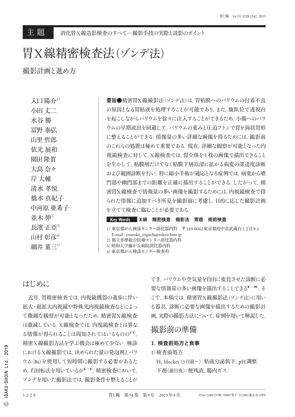
要旨●精密胃X線撮影法(ゾンデ法)は,胃粘膜へのバリウムの付着不良の原因となる胃粘液を処理することが可能であり,また,腹臥位で透視台を起こしながらバリウムを徐々に注入することができるため,小腸へのバリウムの早期流出を回避して,バリウムの重みと圧迫フトンで胃を鉤状胃形に整えることができる.情報量の多い詳細な画像を得るためには,撮影前のこれらの処置は極めて重要である.現在,詳細な観察が可能となった内視鏡検査に対して,X線検査では,胃全体を1枚の画像で描出できることを生かして,粘膜層だけでなく粘膜下層以深に拡がる病変の深達度診断および範囲診断を行い,特に縮小手術が適応となる症例では,病変から噴門部や幽門部までの距離を正確に描出することができる.したがって,精密胃X線検査で情報量の多い画像を撮影するためには,内視鏡検査で得られた情報に追加すべき所見を撮影前に考慮し,目的に応じた撮影計画を立てて検査に臨むことが必要である.
Detailed gastric radiography(Sonde method)enables the processing of gastric mucus, which prevents adhesion of barium to the gastric mucosa. The adhesion of barium facilitates formation of a hook-shaped stomach in the prone position on an upright fluoroscopic table. Such pre-scan processing is extremely important to obtain images with detailed information.
Using radiography, practitioners are able to diagnose and assess the extent of wide-spread lesions and accurately measure the distance from the lesion to the cardia and pylorus. Therefore, it is important to consider the necessary image findings prior to scanning and plan the scan and approach tests in accordance with these goals.

Copyright © 2019, Igaku-Shoin Ltd. All rights reserved.


