Japanese
English
- 有料閲覧
- Abstract 文献概要
- 1ページ目 Look Inside
- 参考文献 Reference
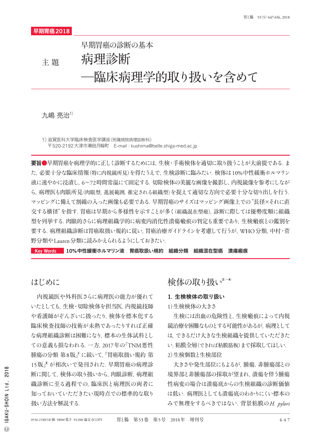
要旨●早期胃癌を病理学的に正しく診断するためには,生検・手術検体を適切に取り扱うことが大前提である.また,必要十分な臨床情報(特に内視鏡所見)を得たうえで,生検診断に臨みたい.検体は10%中性緩衝ホルマリン液に速やかに浸漬し,6〜72時間常温にて固定する.切除検体の美麗な画像を撮影し,内視鏡像を参考にしながら,病理医も肉眼所見(肉眼型,進展範囲,推定される組織型)を捉えて適切な方向で必要十分な切り出しを行う.マッピングに備えて割線の入った画像も必要である.早期胃癌のサイズはマッピング画像上での“長径×それに直交する横径”を指す.胃癌は早期から多様性を示すことが多く(組織混在型癌),診断に際しては優勢度順に組織型を列挙する.肉眼的さらに病理組織学的に病変内消化性潰瘍瘢痕の判定も重要であり,生検瘢痕との鑑別を要する.病理組織診断は胃癌取扱い規約に従い,胃癌治療ガイドラインを考慮して行うが,WHO分類,中村・菅野分類やLauren分類に読みかえられるようにしておきたい.
Although the endoscopists, surgeons, and pathologists are highly skilled, an accurate pathological diagnosis would be difficult and the significance of the specimen as a biological sample could be impaired if the biopsy/resected specimens are seriously treated by clinicians, nurses, or medical technicians. On the other hand, the 8th edition of the TNM classification and the 15th edition of the Japanese Classification of Gastric Carcinoma have been published in 2016 and 2017, respectively. For the pathological diagnosis of early gastric cancer, we explain the standard methods used during the process from tissue sampling to macroscopic and histopathological diagnosis ; both clinicians and pathologists should be aware of these methods.

Copyright © 2018, Igaku-Shoin Ltd. All rights reserved.


