Japanese
English
- 有料閲覧
- Abstract 文献概要
- 1ページ目 Look Inside
- 参考文献 Reference
- サイト内被引用 Cited by
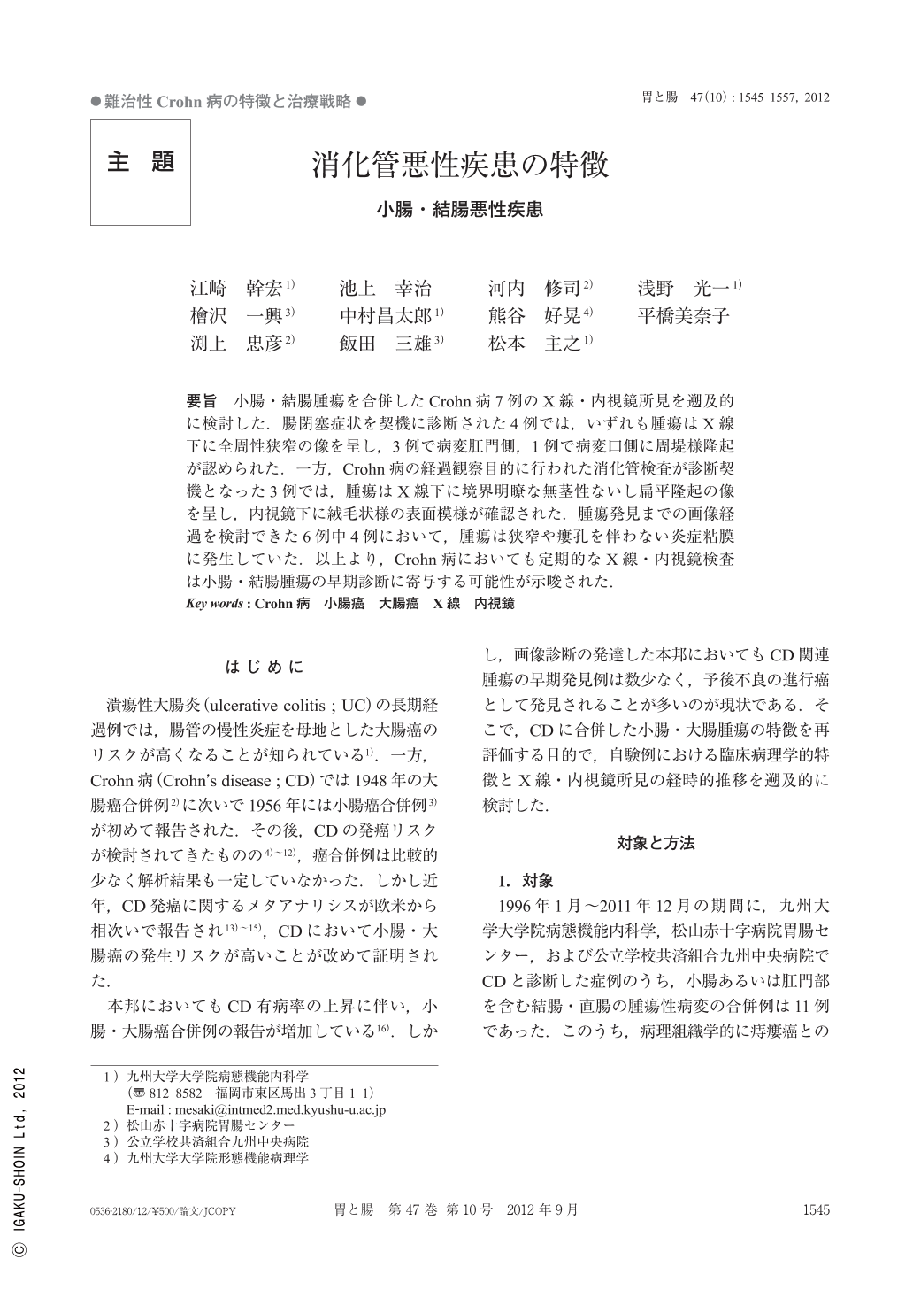
要旨 小腸・結腸腫瘍を合併したCrohn病7例のX線・内視鏡所見を遡及的に検討した.腸閉塞症状を契機に診断された4例では,いずれも腫瘍はX線下に全周性狭窄の像を呈し,3例で病変肛門側,1例で病変口側に周堤様隆起が認められた.一方,Crohn病の経過観察目的に行われた消化管検査が診断契機となった3例では,腫瘍はX線下に境界明瞭な無茎性ないし扁平隆起の像を呈し,内視鏡下に絨毛状様の表面模様が確認された.腫瘍発見までの画像経過を検討できた6例中4例において,腫瘍は狭窄や瘻孔を伴わない炎症粘膜に発生していた.以上より,Crohn病においても定期的なX線・内視鏡検査は小腸・結腸腫瘍の早期診断に寄与する可能性が示唆された.
We retrospectively investigated endoscopic and radiographic characteristics of 7patients with CD(Crohn's disease)complicated by cancer of the small or large intestine. At the time of diagnosis, 4patients manifested nausea, vomiting, and abdominal colicky pain, suggesting severe intestinal stenosis. Subsequent radiography depicted occlusive luminal stenosis mimicking the inflammatory luminal stenosis of CD. However, the tumor edge was demonstrated at the anal side of the stenosis in 3patients, and at the oral side in 1patient. On the other hand, follow-up GI examination for luminal CD revealed complicating cancer in the other 3patients. They complained of no cancer related abdominal symptoms. Radiographically, the tumor was depicted as a sessile or flat protrusion, in which villous like surface was observed under endoscopy. Among those 6patients, the tumor had arisen from chronic inflammatory mucosa in 4patients. It thus can be suggested that periodical GI examination is mandatory for early detection of complicating cancer, as well as for the evaluation of mucosal lesions of CD.

Copyright © 2012, Igaku-Shoin Ltd. All rights reserved.


