Japanese
English
- 有料閲覧
- Abstract 文献概要
- 1ページ目 Look Inside
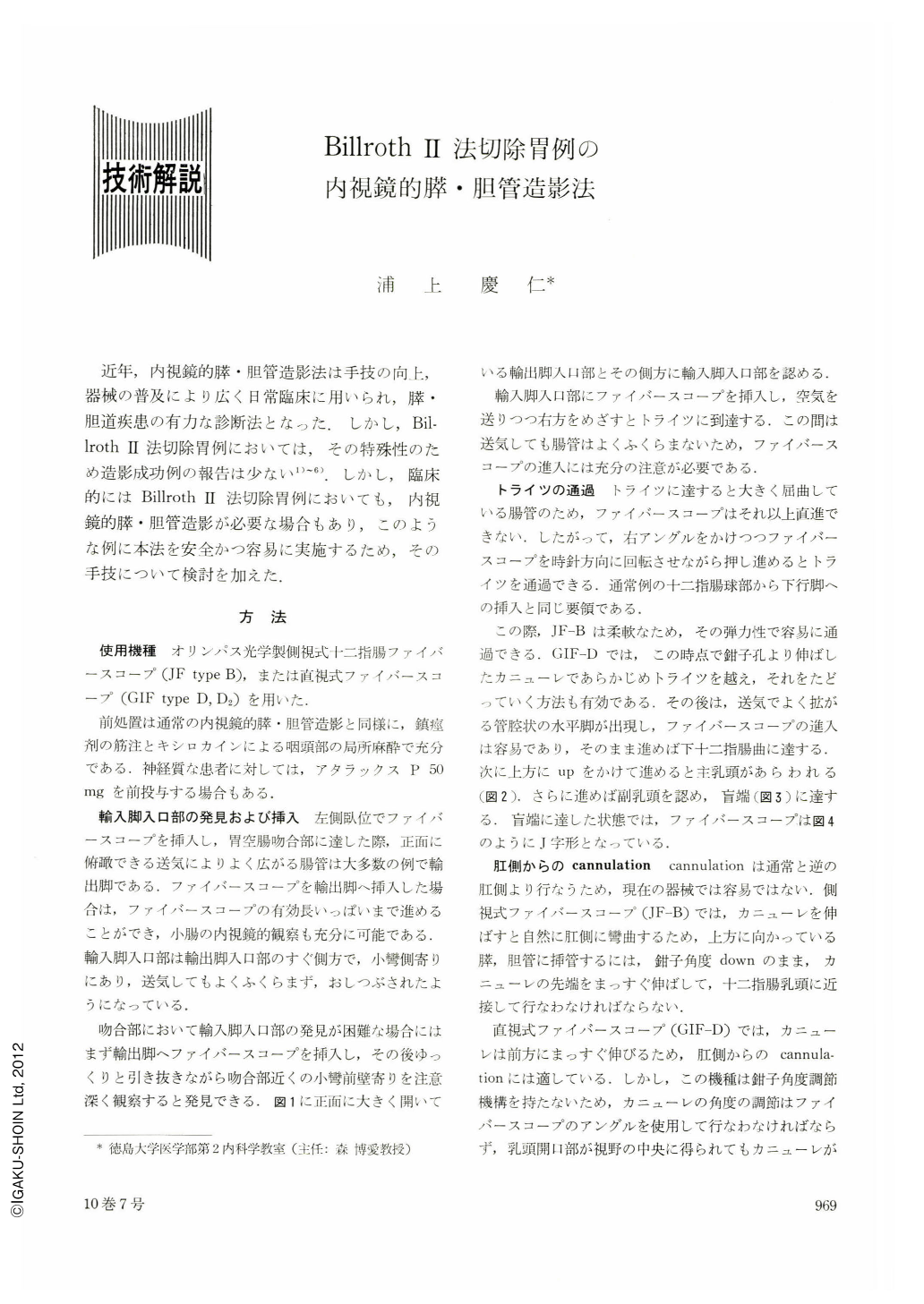
近年,内視鏡的膵・胆管造影法は手技の向上,器械の普及により広く日常臨床に用いられ,膵・胆道疾患の有力な診断法となった.しかし,Billroth Ⅱ法切除胃例においては,その特殊性のため造影成功例の報告は少ない1)~6).しかし,臨床的にはBillroth Ⅱ法切除胃例においても,内視鏡的膵・胆管造影が必要な場合もあり,このような例に本法を安全かつ容易に実施するため,その手技について検討を加えた.
In recent years endoscopic pancreatocholangiography has become a most effective diagnostic method for diseases of the pancreas and bile duct. However, in patients gastrectomized according to Billroth II method, reports are few in the successful depiction of these parts. A study has therefore been made in the endoscopic exposure of the pancreas and bile duct in patients whose stomachs were resected according to Billroth II method, by using JF-B, a side-viewing fiberscope, and GIF-D, a forward-viewing fiberscope.
Of 25 such cases examined by these instruments, 20 (80%) were successfully depicted, 7 cases by JF-B and 13 by GIF-D. The pancreatic duct was visualized by JF-B in all of the 7 cases (100%) and the bile duct in 3 (43%). By GIF-D the pancreatic duct was successfully exposed in 11 cases (85%) and the bile duct in 8 (62%). Both ducts were visualized by JF-B in 3 cases (43%) and by GIF-D in 6 (46%). As with the ordinary cases indicated for pancreatocholangiography, the pancreatic duct in gastrectomized cases according to Billroth II method was more easily visualized than the bile duct by both side-viewing and forward-viewing fiberscopes. The cannula of the JF-B would bend down at the time of cannulation in such a manner as to make the pancreatic duct radiopaque far easier than the bile duct. On the other hand, GIF-D showed a higher positive rate in the depiction of the bile duct because its cannula would come out straight ahead.
Of fiberscopes available at present, it would be advisable thus to select a side-viewing fiberscope for pancreatography and a forward-viewing one for cholangiography. The rate of success would be intensified in pancreatocholangiography if a forward-viewing fiberscope be used equipped with a cannula having adjustable angle device. Technically, the following three points are most difficult to manage: (1) detection and cannulation of the orifice in the afferent loop; (2) passage of fiberscope through the segment at the level of the Treitz' ligament; (3)and cannulation from the anal side. These techniques have been explained here.
The present method is believed to ensure effective endoscopic approach to postgastrectomy syndromes as well as to afferent loop syndrome.

Copyright © 1975, Igaku-Shoin Ltd. All rights reserved.


