Japanese
English
- 有料閲覧
- Abstract 文献概要
- 1ページ目 Look Inside
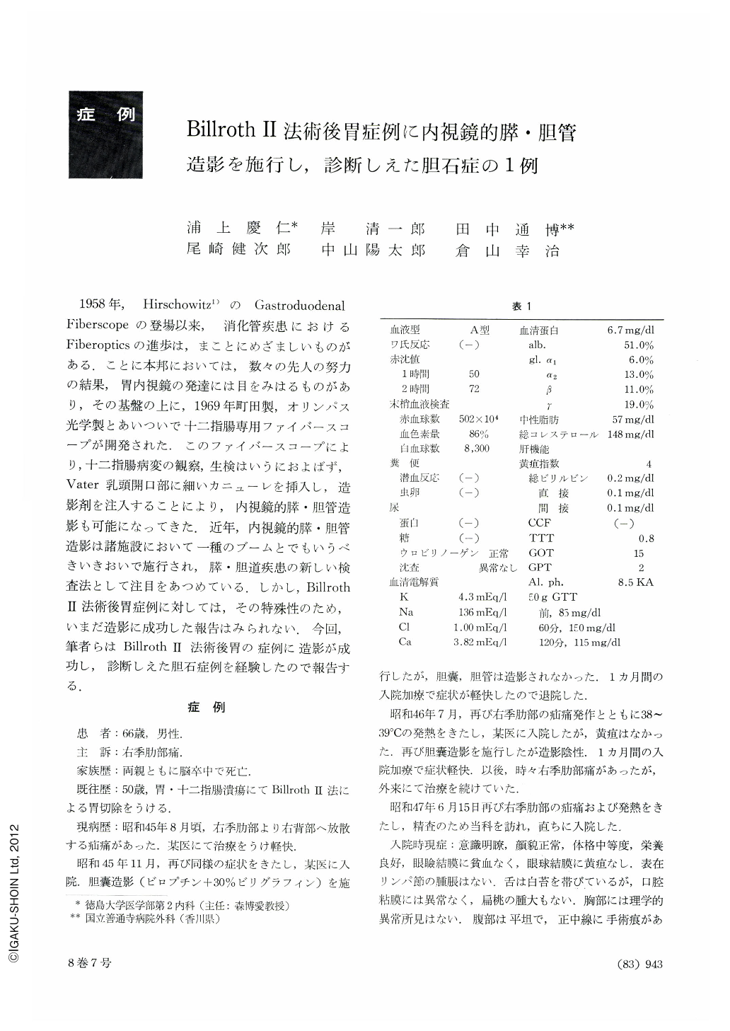
1958年,Hirschowitz1)のGastroduodenal Fiberscopeの登場以来,消化管疾患におけるFiberopticsの進歩は,まことにめざましいものがある.ことに本邦においては,数々の先人の努力の結果,胃内視鏡の発達には目をみはるものがあり,その基盤の上に,1969年町田製,オリンパス光学製とあいついで十二指腸専用ファイバースコープが開発された.このファイバースコープにより,十二指腸病変の観察,生検はいうにおよぼず,Vater乳頭開口部に細いカニューレを挿入し,造影剤を注入することにより,内視鏡的膵・胆管造影も可能になってきた.近年,内視鏡的膵・胆管造影は諸施設において一種のブームとでもいうべきいきおいで施行され,膵・胆道疾患の新しい検査法として注目をあつめている.しかし,Billroth Ⅱ法術後胃症例に対しては,その特殊性のため,いまだ造影に成功した報告はみられない.今回,筆者らはBillroth Ⅱ法術後胃の症例に造影が成功し,診断しえた胆石症例を経験したので報告する.
A man aged 66 had undergone gastrectomy (Billroth II) 16 years before on account of gastroduodenal ulcer. Since about August 1970 he had bouts of colic, several times a year, in the right hypochondrium, radiating to the right-hand side of the back. In June 1972 he was admitted to our hospital for thorough check up. Upper gastrointestinal x-ray series showed that gastrectomy had been done according to the Billroth II method. It also revealed the duodenal papilla and accesory papilla clearly visualized in the afferent loop. Combined peroral and intravenous cholecystocholangiography disclosed the dilated common bile duct, with the gallbladder remaining unopacified. Endoscopic pancreatocholangiography showed normal pancreatic ducts, revealing not only the common bile duct but hepatic ducts as well. The cystic duct was also visualized, but still the gallbladder failed to become radiopaque. Subsequent surgical intervention showed a very atro-phic gallbladder with a thickened wall. A septum was found in the fundus, with a stone beneath it.
In recent years duodenofoberscopy has received sucha wide acceptance that endoscopic pancreatocholangiography is now performed in various institutions, its results being reported one after another. According to Ogoshi et al., positive rate of visualization is as high as 96.4 per cent. Tremendous as has been the progress made in the field of pancreatocholangiography, the afferent loop in patients gastrectomized according to Billroth II method is still out of reach for this procedure. The afferent loop spot still remains the blind spot. So far not a single report with positive result has been encountered.
The instrument employed in the present study is the duodenofiberscope JF type B (Olympus).

Copyright © 1973, Igaku-Shoin Ltd. All rights reserved.


