Japanese
English
- 有料閲覧
- Abstract 文献概要
- 1ページ目 Look Inside
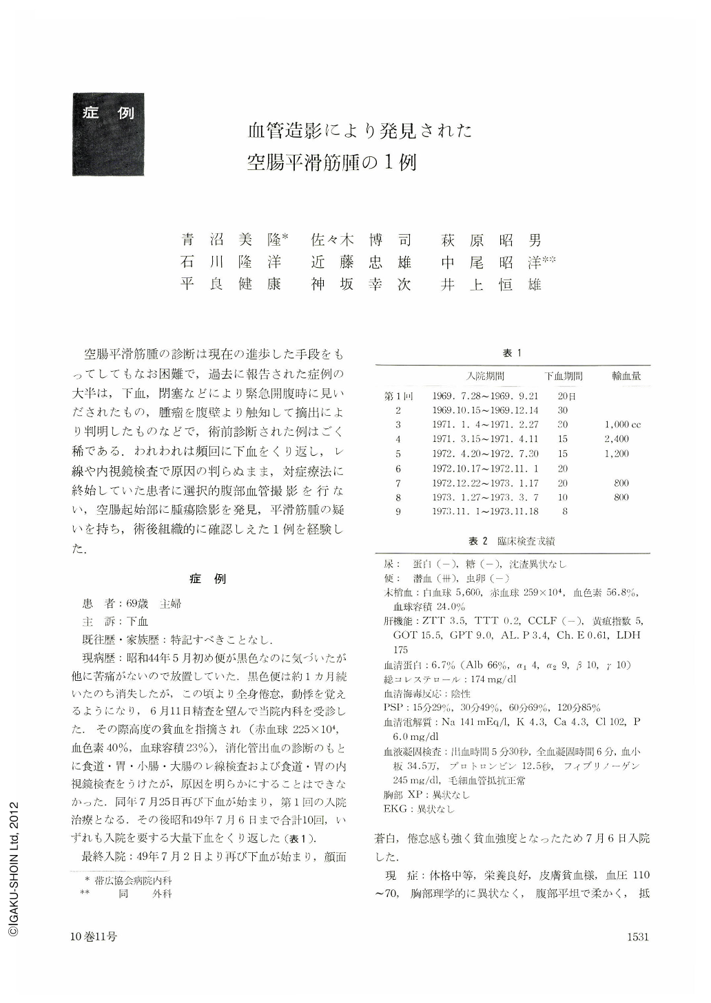
空腸平滑筋腫の診断は現在の進歩した手段をもってしてもなお困難で,過去に報告された症例の大半は,下血,閉塞などにより緊急開腹時に見いだされたもの,腫瘤を腹壁より触知して摘出により判明したものなどで,術前診断された例はごく稀である.われわれは頻回に下血をくり返し,レ線や内視鏡検査で原因の判らぬまま,対症療法に終始していた患者に選択的腹部血管撮影を行ない,空腸起始部に腫瘍陰影を発見,平滑筋腫の疑いを持ち,術後組織的に確認しえた1例を経験した.
This is a case preoperatively diagnosed as a tumor of the jejunum by angiography and was confirmed as a Leiomyoma of jejunum by histological examination.
A 69-year-oId female patient noticed tarry stool in the beginning of May 1969. It persisted for one month and then subsided. On June 11, 1969, she visited our hospital with a chief complaint of weakness. On physical examination she was found to be moderately anemic, but had no other abnormal signs, and no abdominal mass was palpated. The routine X-ray examination showed no abnormal change in the upper and lower digestive tract. No abnormality was seen by the endoscopic examination of the esophagus and stomach.
During the subsequent 5 years from the initial bleeding to July 1974, when finally she was admitted to our hospital, she had nine attacks of bloody discharge that lasted for one week to one month. Whenever bleeding occurred, she entered our hospital and received supplemental treatment with many blood transfusions for anemia. Repeatedly the X-ray and endoscopic examinations of the digestive tract were tried, but no abnormality was found. On her final entrance in August 1974, selective angiography of the abdominal cavity showed a small mass at the orifice of the jejunum.
Retrospective study of this case revealed that the X-ray film taken in June 1969 showed a round-like defect in the orifice of the jejunum.
Surgical extirpation showed an oval-shaped tumor developing mostly outside of the jejunum wall, about 7 cm anal from Treitz-ligament. Neither infiltration nor metastasis was found in the adjoining areas or in abdominal lymph nodes. The tumor size was about 1.5×1.5×2 cms. Histologically it was diagnosed as a benign leiomyoma. Finally, it is especially emphasized here that selective abdominal angiography was the most effective diagnostic procedure in this case.

Copyright © 1975, Igaku-Shoin Ltd. All rights reserved.


