Japanese
English
- 有料閲覧
- Abstract 文献概要
- 1ページ目 Look Inside
- サイト内被引用 Cited by
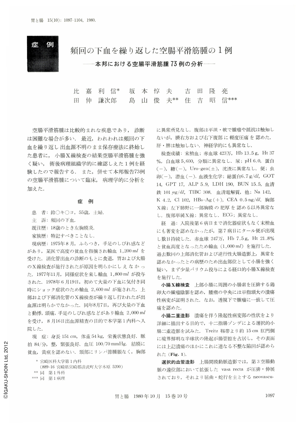
空腸平滑筋腫は比較的まれな疾患であり,診断は困難な場合が多い.最近,われわれは頻回の下血を繰り返し出血源不明のまま保存療法に終始した患者に,小腸X線検査の結果空腸平滑筋腫を強く疑い,術後病理組織学的に確認しえた1例を経験したので報告する.また,併せて本邦報告73例の空腸平滑筋腫について臨床,病理学的に分析を加えた.
A 55 year-old woman has had episodes of recurrent gastrointestinal bleeding for three years. Several routine x-ray examinations showed no abnormality in the upper and lower digestive tracts. A barium follow through examination of the small intestine disclosed a round mass with central ulceration at the proximal jejunum, which was subsequently more precisely delineated by a double contrast study of the small intestine and by a selective angiography of the superior mesenteric artery.
The patient was operated on under a presumptive diagnosis of leiomyoma. Surgical specimen showed a hypervascular tumor developing both inside and outside the intestinal lumen, 17 cm distal to the ligament of Treitz. There was neither infiltration nor metastasis in the adjacent organ except for enlargement of several regional lymph nodes. The tumor excised revealed a “dumbbell” shaped growth, the small endoenteric portion measuring 4.2×3.8×2.7 cm and the larger exoenteric portion measuring 6.0×4.0×3.8 cm. There was an irregular ulceration extending into the tumor and developing into a cystic cavity filled with hemorrhagic necrosis. Histologically, it was diagnosed as a benign leiomyoma, in part of which mitoses were found.
A total of 73 cases of leiomyoma of the jejunum has been reported from 1912 to 1978 in Japan. Cardinal symptoms noted were gastrointestinal bleeding and anemia 61.2%, abdominal pain 43.3%, and palpable mass 37.3%. Exoenteric growth was observed in 67.2%, endoenteric in 15.6%, and mixed in 17.2%. In a majority of the cases (67.2%), the size of the tumor was more than 5 cm in the maximum diameter. Many of exoenteric tumors undergo degenerative changes such as ulceration, central necrosis, or pseudocystic alteration. This tendency was found more in the larger tumors. Seven cases of leiomyoma with perforation were all confined to the exoenteric group.

Copyright © 1980, Igaku-Shoin Ltd. All rights reserved.


