Japanese
English
- 有料閲覧
- Abstract 文献概要
- 1ページ目 Look Inside
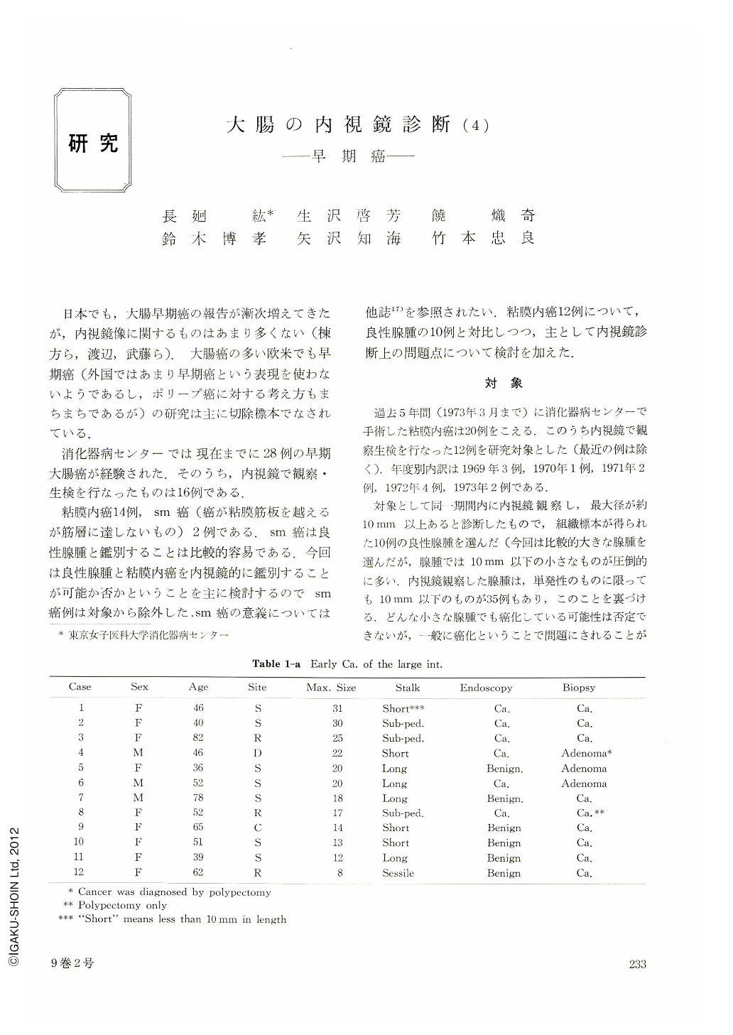
日本でも,大腸早期癌の報告が漸次増えてきたが,内視鏡像に関するものはあまり多くない(棟方ら,渡辺,武藤ら).大腸癌の多い欧米でも早期癌(外国ではあまり早期癌という表現を使わないようであるし,ポリープ癌に対する考え方もまちまちであるが)の研究は主に切除標本でなされている.
消化器病センターでは現在までに28例の早期大腸癌が経験された.そのうち,内視鏡で観察・生検を行なったものは16例である.
Twelve cases of cancer in adenoma (early cancer) and eleven cases of benign adenoma were selected for this study. All of them were observed endoscopically and entire specimen could be gained for histological examination. Serial block sections were carefully examined. Cases of polyposis were excluded from this survey because of the difficulty in thorough examination. Average age of patients with early cancer was 54, ranging from 21 to 69. It was revealed that the invasion did not exceed the muscularis mucosae in all of our early cancers. There is no significant morphological difference between malignant and benign adenoma. The mean maximal size of 12 early cancers was 19 mm (8~31 mm), and 14 mm (8~27 mm) in the benign adenomas.
There is evident overlapping in size of benign and malignant adenoma. It is generally agreed that the larger the adenoma the higher the risk of malignant change. In each individual adenoma, however, we can not say its nature according to its size alone. The morphologic characteristic of the early cancers in this series were as follows; pedunculated, 8 cases; sub-pedunculated, three; sessile, one case. Those of the benign adenoma were; pedunculated, 10; sessile 1.
It was also impossible to know whether the adenoma is benign or malignant from its gross appearances. Surface nodularity seems to reflect the size of the tumor, and it cannot be relied as an indicator of malignant change. It was also found that erosion and redness of the head were not so reliable change as for the nature of the tumor. The large intestine often suffers from mechanical stimulation from hard faces etc., so the tumors easily get eroded. The role of biopsy under direct vision is great in the diagnosis of tumors of large intestine; Colonoscope permits to take biopsy specimens at any parts of the large intestine. It is often difficult, however, to take biopsy from a pedunculated tumor. When the biopsy forcep touchs the head of the polyp it leans towards opposite direction. Usually we take two to five specimens from the head of the tumor and one or two from the stalk of the pedunculated polyp. Biopsy was negative in three cases (case 4. 5. 6) of early cancer, all of which were pedunculated. In case 4, carcinoma was restricted and surface area showed no malignancy. Therefore, it seemed impossible to diagnose carcinoma by biopsy in this case. In case 5, carcinoma was found only at the tip of the tumor. Satisfactory specimen could not be gained from that part. In case 6, almost entire surface area showed carcinomatous change. Histological examination of biopsy material revealed atypism.
It was impossible to conclude the tumor to be malignant from the material.
Negative biopsy does not rule out the malignancy, especially in cases of pedunculated tumor. It is possible to polypectomize any pedunculated polyp of the large intestine. Endoscopic removal of colonic polyps is now proposed as a safe, practical alternative to either laparotomy and colotomy or repeated barium-enema studies in the management of the patient with a colonic polyp. Sometimes endoscopic diagnosis of early cancer was not sure. Even biopsy under direct vision did not always give us true information. Hence, polypectomy can be said to be the sole complete diagnostic procedure.

Copyright © 1974, Igaku-Shoin Ltd. All rights reserved.


