Japanese
English
- 有料閲覧
PPV購入案内
1,320円 (1,200円+税10%) こちらは、789ページから794ページの文献です。購入には医書.jp本会員登録が必要です。
会員登録完了後に改めて上記「購入サイトへ」を選択してください。
または「購入サイトへ」選択後に「購入ログイン」、「新規会員登録」の順に進んでください。
会員登録について詳しくはをご確認ください。
- Abstract 文献概要
- 1ページ目 Look Inside
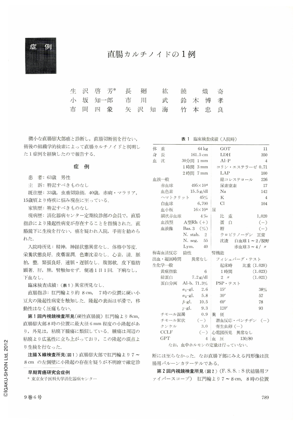
微小な直腸膨大部癌と診断し,直腸切断術を行ない,術後の組織学的検索によって直腸カルチノイドと判明した1症例を経験したので報告する.
患 者:63歳 男性
主 訴:特記すべきものなし
既往歴:33歳,虫垂切除術.40歳,赤痢・マラリア,15歳頃より痔疾に悩み現在に至っている.
家族歴:特記するべきものなし
現病歴:消化器病センター定期検診部の会員で,直腸指診により隆起性病変が存在することを指摘された.直腸鏡下に生検を行ない,癌を疑われ入院,手術を勧められた.
A case of rectal carcinoid, whose histological differentiation from adenocarcinoma was very difficult, was correctly diagnosed by electronmicroscopy.
A patient, 63 years old male, consulted our hospital for periodic health examination. He complained of no lower abdominal symptoms. Digital examination of the rectum revealed a small hard nodule. Endoscopically the tiny nodule was of yellowish cast and the overlying mucosa was almost normal. Its diameter was assumed to be about 9 mm. No ulceration and erosion were observed.
Opetation was carried out because of the difficulty of the differentiation of carcinoid from adenocarcinoma. Silver stains were negative. Electron microscopic examination of the resected specimen revealed numerous dense particles, characteristic for carcinoid tumor. The diagnosis of carcinoid tumor was then established.
Copyright © 1974, Igaku-Shoin Ltd. All rights reserved.