Japanese
English
- 有料閲覧
- Abstract 文献概要
- 1ページ目 Look Inside
潰瘍性大腸炎が文献にみられるようになったのは19世紀も後半になってからである.最初の報告としてよく引用されるのはWilks1)の論文である.さらにさかのぼって探そうとして諸種の論文が挙げてあるが,現在でも未解決の点の多い疾患であるので不備な古い論文からあまり確実なことはいえないと思う.いずれにしてもそう古くない疾患であるといえる.しかし潰瘍性大腸炎は,現在では,少くとも欧米においては最もポピュラーな大腸疾患の一つである.日本においても戦後漸増し,大腸の感染症が減少した今日では,下血をきたす患者において癌,ポリープと並んで先ず考慮すべき疾患となっている.消化器病センターでも過去約2年半の間に,ファイバースコープで観察した症例だけに限っても34例に達している.今後益々増えることが予想される.この論文では内視鏡像と生検組織に重点をおいて,潰瘍性大腸炎について述べる.
材料
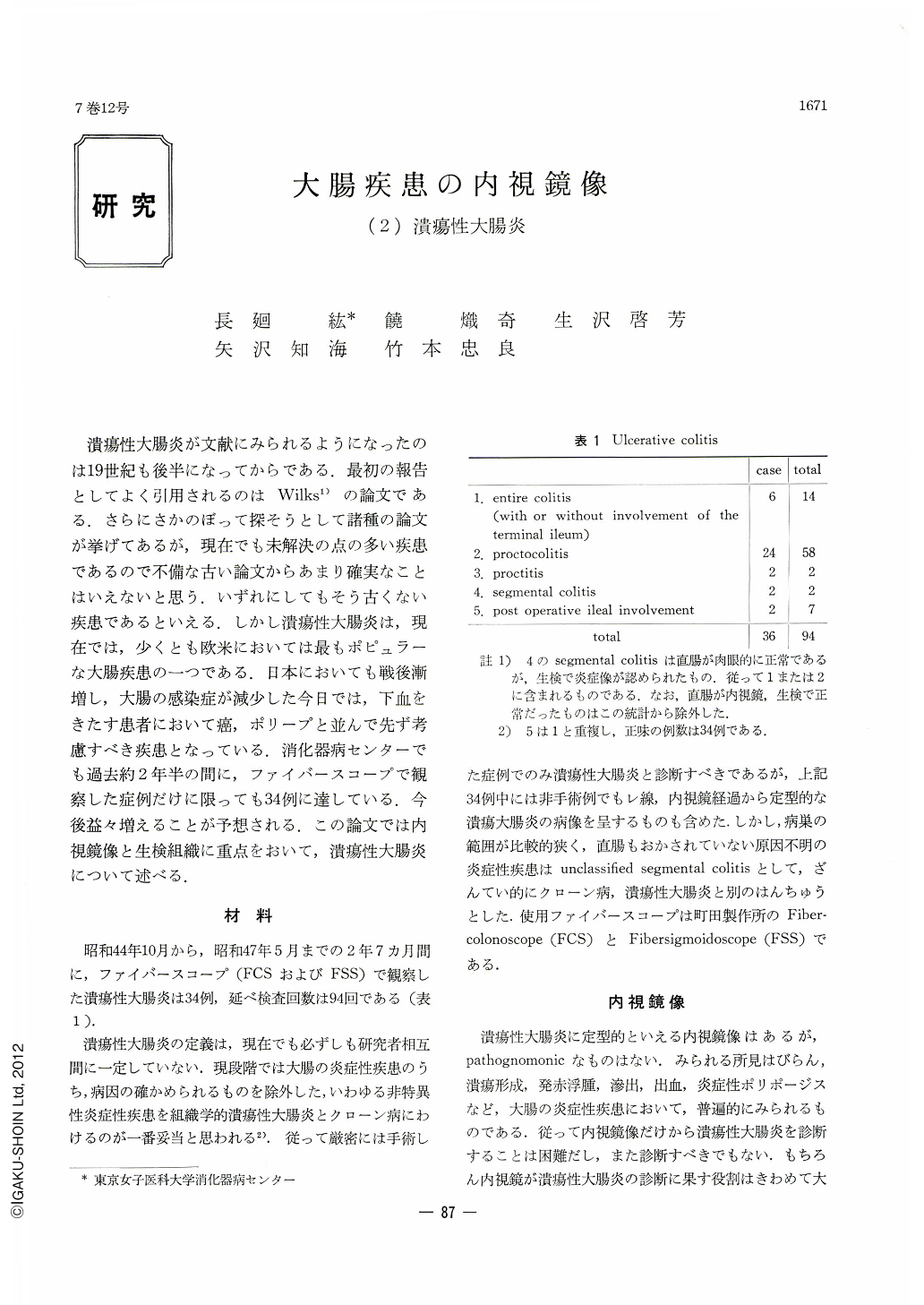
昭和44年10月から,昭和47年5月までの2年7カ月間に,ファイバースコープ(FCSおよびFSS)で観察した潰瘍性大腸炎は34例,延べ検査回数は94回である(表1).
During the two years and seven months from Oct. 1969 to May 1972 ulcerative colitis was observed by fiberscope (FCS and FSS) in 34 patients, totalling to 94 examinations. Ulcerative colitis, representing unspecific inflammatory disease of the colon other than Crohns' disease or unclassified (segmental) colitis, was in the past mostly examined by hard-type rectoscope, with its merits properly evaluated. Introduction of fiberscope in the field of colon endoscope has now broadened the extent of macroscopic examination in diseases of the large intestine. Endoscopically we have divided ulcerative colitis into acute and chronic stages, and the former further into slight, moderate and severe degrees, while the latter was divided into active and inactive stages. Needless to say, every stage of the disease is not always possible to distinguish in an exact manner. Inflammatory ulcer formation seen in ulcerative colitis is characterized by shallow ulcers of irregular shape. They become confluent, forming larger ulcers. There are islet-like mucosal residues in between. However, not infrequently no ulcer formation can be seen. Ulcers tend to undermine the surface and to form mucosal tags, giving birth to inflammatory polyps. They present very varied pictures as they merge into one another. when the rectum may look normal in ulcerative colitis, biopsy would often demonstrate inflammation. Contrary to Crohn's disease, ulcerative colitis is characterized by the uniformity of its pathologic pictures.
Ulcerative colitis shows no pathognomonic histological pictures. Biopsy has its limitations, and yet it can be of great help in case endoscopy fails to reveal inflammation, or when the degree of inflammation remains uncertain. Accordingly, one should collect biopsy specimens from variuos parts of different segments. Biopsy is more dependable than endoscopy when one has to establish the extent of inflammation in ulcerative colitis.

Copyright © 1972, Igaku-Shoin Ltd. All rights reserved.


