Japanese
English
- 有料閲覧
- Abstract 文献概要
- 1ページ目 Look Inside
- サイト内被引用 Cited by
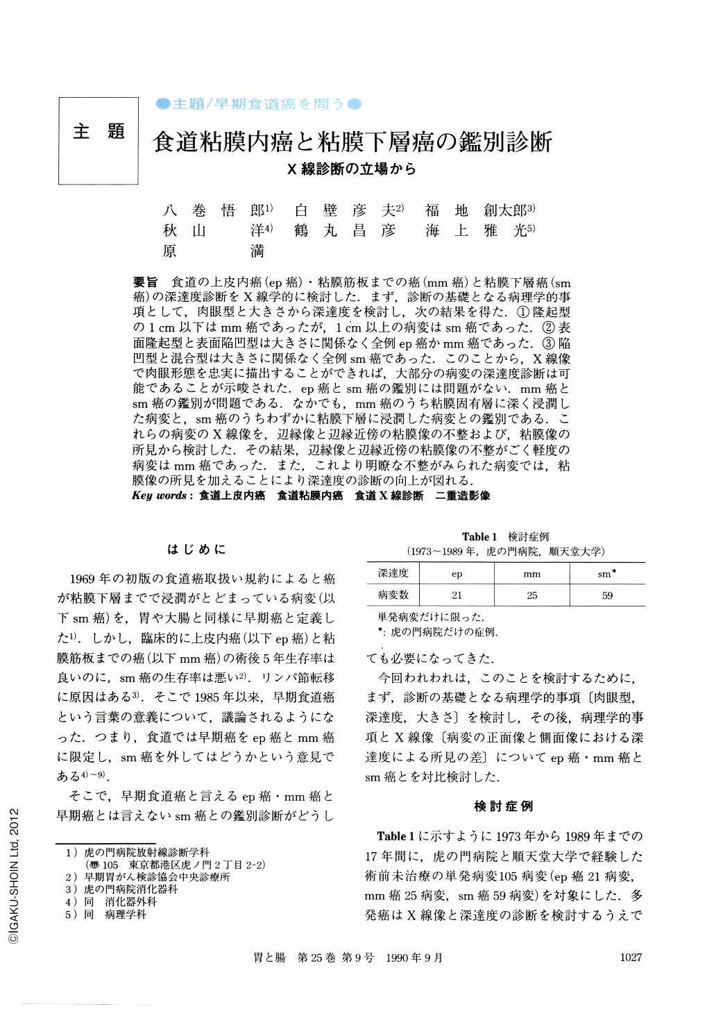
要旨 食道の上皮内癌(ep癌)・粘膜筋板までの癌(mm癌)と粘膜下層癌(sm癌)の深達度診断をX線学的に検討した.まず,診断の基礎となる病理学的事項として,肉眼型と大きさから深達度を検討し,次の結果を得た.①隆起型の1cm以下はmm癌であったが,1cm以上の病変はsm癌であった.②表面隆起型と表面陥凹型は大きさに関係なく全例ep癌かmm癌であった.③陥凹型と混合型は大きさに関係なく全例sm癌であった.このことから,X線像で肉眼形態を忠実に描出することができれば,大部分の病変の深達度診断は可能であることが示唆された.ep癌とsm癌の鑑別には問題がない.mm癌とsm癌の鑑別が問題である.なかでも,mm癌のうち粘膜固有層に深く浸潤した病変と,sm癌のうちわずかに粘膜下層に浸潤した病変との鑑別である.これらの病変のX線像を,辺縁像と辺縁近傍の粘膜像の不整および,粘膜像の所見から検討した.その結果,辺縁像と辺縁近傍の粘膜像の不整がごく軽度の病変はmm癌であった.また,これより明瞭な不整がみられた病変では,粘膜像の所見を加えることにより深達度の診断の向上が図れる.
In the last seventeen years, twenty-one cases with single lesion of intraepithelial carcinoma (ep-ca.), 25 cases with single lesion of mucosal carcinoma (mm-ca.) and 59 cases with single lesion of submucosal carcinoma (sm-ca.) have been detected and resected in our hospital.
The gross findings of ep-ca. and mm-ca. were classified into four basic types of early gastric cancer (Type Ⅰ, Ⅱa, Ⅱb and Ⅱc). On the other hand, those of sm-ca. were classified into four types (Type 1, I-like, 2, and Mixed).
In elevated lesions larger than 1 cm, the carcinoma had invaded the submucosa. However, in superficial elevated type lesions, the carcinomas were confined to the intramucosa. This confinement of the cancerous invasion to the intramucosa was also found in superficial depressed type lesions.
On the other hand, regardless of their size, in depressed and mixed-type lesions, the carcinoma always invaded the submucosa.
So, it is possible for us to diagnose the depth of cancerous invasion, if we depict the lesion faithfully in the double contrast pictures.
In radiological study, it is easy to locate the lesions with distinguished elevation or depression, but it is difficult to depict the lesions accompanied by epithelial invasion. We must interpret the irregularity of the esophageal wall first, and then interpret the irregularity of the mucosal pattern.
Copyright © 1990, Igaku-Shoin Ltd. All rights reserved.