Japanese
English
- 有料閲覧
- Abstract 文献概要
- 1ページ目 Look Inside
- サイト内被引用 Cited by
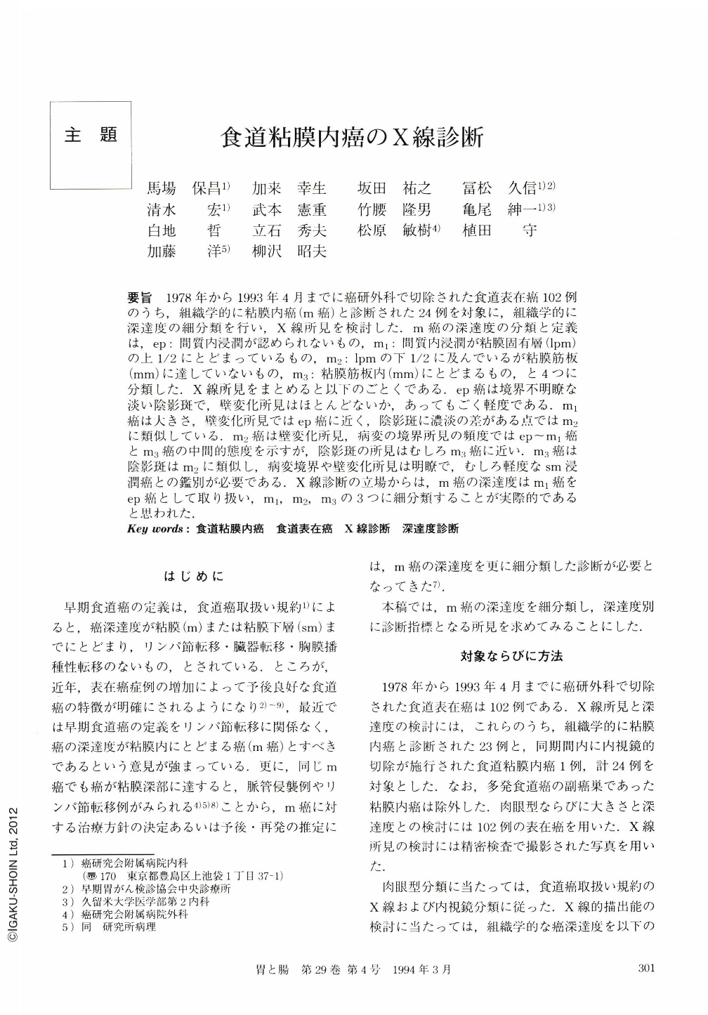
要旨 1978年から1993年4月までに癌研外科で切除された食道表在癌102例のうち,組織学的に粘膜内癌(m癌)と診断された24例を対象に,組織学的に深達度の細分類を行い,X線所見を検討した.m癌の深達度の分類と定義は,ep:間質内浸潤が認められないもの,m1:間質内浸潤が粘膜固有層(lpm)の上1/2にとどまっているもの,m2:lpmの下1/2に及んでいるが粘膜筋板(mm)に達していないもの,m3:粘膜筋板内(mm)にとどまるもの,と4つに分類した.X線所見をまとめると以下のごとくである.ep癌は境界不明瞭な淡い陰影斑で,壁変化所見はほとんどないか,あってもごく軽度である.m1癌は大きさ,壁変化所見ではep癌に近く,陰影斑に濃淡の差がある点ではm2に類似している.m2癌は壁変化所見,病変の境界所見の頻度ではep~m1癌とm3癌の中間的態度を示すが,陰影斑の所見はむしろm3癌に近い.m3癌は陰影斑はm2に類似し,病変境界や壁変化所見は明瞭で,むしろ軽度なsm浸潤癌との鑑別が必要である.X線診断の立場からは,m癌の深達度はm1癌をep癌として取り扱い,m1,m2,m3の3つに細分類することが実際的であると思われた.
We evaluated x-ray findings of 24 superficial esophageal cancers subclassified according to the depth of cancer invasion as follows: ep; those confined to the epithelium, m1; those limited within the upper half of the lamina propria mucosae (lpm), m2; those invading the lower half of lpm, and m3; those involving the lamina muscularis mucosae. On the x-ray examination, ep cancers were visualized as faintest barium shadows with unclear border, and marginal irregularity was very hard to point out. Density of barium shadow became uneven in cases of m1 cancer, and marginal irregularity and the border of lesion became clearer as cancer invades deeper layer. From the radiographical point of view, it is useful to regard ep and m1 cancers as one category, and subclassify esophageal intramucosal cancer as m1, m2, m3.

Copyright © 1994, Igaku-Shoin Ltd. All rights reserved.


