Japanese
English
- 有料閲覧
- Abstract 文献概要
- 1ページ目 Look Inside
- サイト内被引用 Cited by
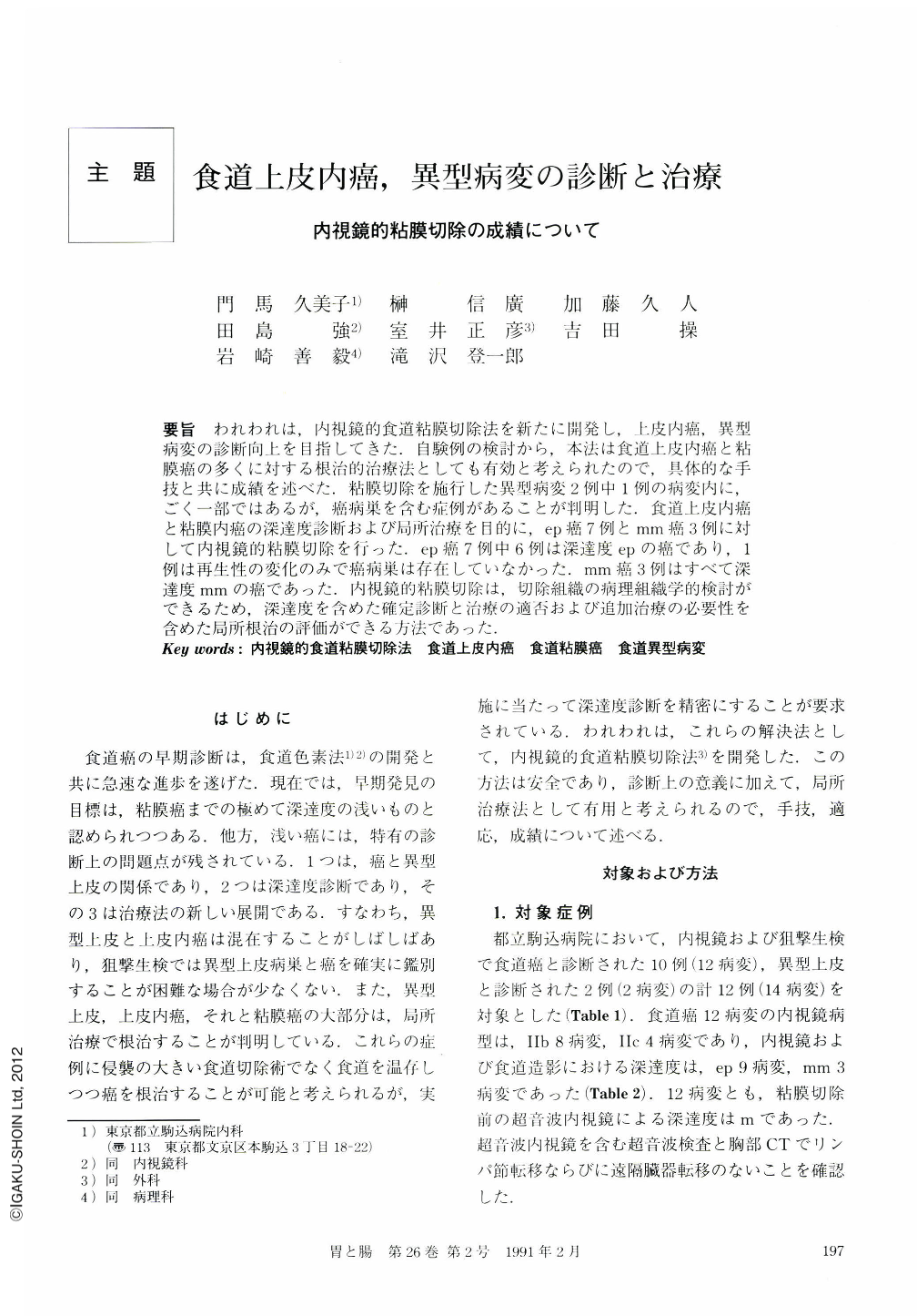
要旨 われわれは,内視鏡的食道粘膜切除法を新たに開発し,上皮内癌,異型病変の診断向上を目指してきた.自験例の検討から,本法は食道上皮内癌と粘膜癌の多くに対する根治的治療法としても有効と考えられたので,具体的な手技と共に成績を述べた.粘膜切除を施行した異型病変2例中1例の病変内に,ごく一部ではあるが,癌病巣を含む症例があることが判明した.食道上皮内癌と粘膜内癌の深達度診断および局所治療を目的に,ep癌7例とmm癌3例に対して内視鏡的粘膜切除を行った.ep癌7例中6例は深達度epの癌であり,1例は再生性の変化のみで癌病巣は存在していなかった.mm癌3例はすべて深達度mmの癌であった.内視鏡的粘膜切除は,切除組織の病理組織学的検討ができるため,深達度を含めた確定診断と治療の適否および追加治療の必要性を含めた局所根治の評価ができる方法であった.
Lymph node metastasis is rare in intraepithelial or mucosal squamous cell carcinoma of the esophagus. An excision of the mucosa including the lesion may be curative for it. Endoscopic mucosectomy technique allows us to remove the mucosa and the submucosa including the lesion preserving the proper muscle layer.We used this technique in precisely predicting the presence of and treating mucosal carcinoma of the esophagus.
Endoscopic mucosectomy was carried out in 12 cases with esophageal mucosal lesions: tentative diagnosis were intraepithelial cancer in 7 cases, mucosal cancer in 3 and atypical epithelium 2 before the procedure. Lymph node metastasis was not suspected by CT-scan, endoscopic ultrasonography or conventional ultrasonography.
Results obtained are as follows:
1) Size of the removed mucosa: The largest dimension of the piece of the resected mucosa was approximately 15 mm, indicating that the maximal dimension of a lesion removable by single procedure of resection was approximately 12 mm.
2) Completeness of resection: A lesion with dimension over 12 mm could be removed by repeating mucosectomy several times. Mucosectomy was done first from the central portion, and then peripheral portion with the surrounding normal mucosa. Endoscopic staining technique using iodine facilitated the procedure. When a lesion was removed without leaving any small islands of cancerous tissue, the procedure was regarded as complete resection. In 11 out of 12 cases resection was regarded as complete and the remaining one incomplete.
3) No recurrence was observed in cases with complete resection but it occurred in a case with imcomplete resection 6 months later. That case was immediately treated surgically. An intraepithelial carcinoma was found in the resected specimen. Therefore, endoscopic examination was recommended 3, 6, and 12 months after endoscopic mucosectomy for at least one year.
4) Preoperative diagnoses of mucosal lesion and pathological studies on mucosectomy specimens: Squamous cell carcinoma was detected in 10 cases before the procedure and confirmed by mucosectomy. Intraepithelial carcinoma was not detected until bite biopsy was done before the mucosectomy in 1 case. Degenerated epithelium was wide-spread in the lesion. In 1 of the 2 cases with atypical epithelial lesions, a small focal squamous cell carcinoma was found in the basal layer by endoscopic mucosectomy. Intraepithelial carcinoma was correctly diagnosed in 7 cases before the treatment and mucosal cancer in 3 cases. One of the 2 atypical epithelial cases had intraepithelial carcinoma and the other, atypical epithelial change.
Thus, it is imperative that we consider on the fact that atypical epithelial lesion of the esophagus is likely to have small carcinoma in it. It is possible to pathologically examine in detail large mucosal pieces obtained by endoscopic mucosectomy. Depth of ivasion of intraepithelial and mucosal carcinoma of the esophagus is correctly and reliably evaluated by conventional endoscopy and bite biopsy. It is feasible to remove the lesion completely by endoscopic mucosectomy. When a single procedure of endoscopic mucosectomy is considered as complete, we may judge it a curative treatment.
Copyright © 1991, Igaku-Shoin Ltd. All rights reserved.