Japanese
English
- 有料閲覧
- Abstract 文献概要
- 1ページ目 Look Inside
- サイト内被引用 Cited by
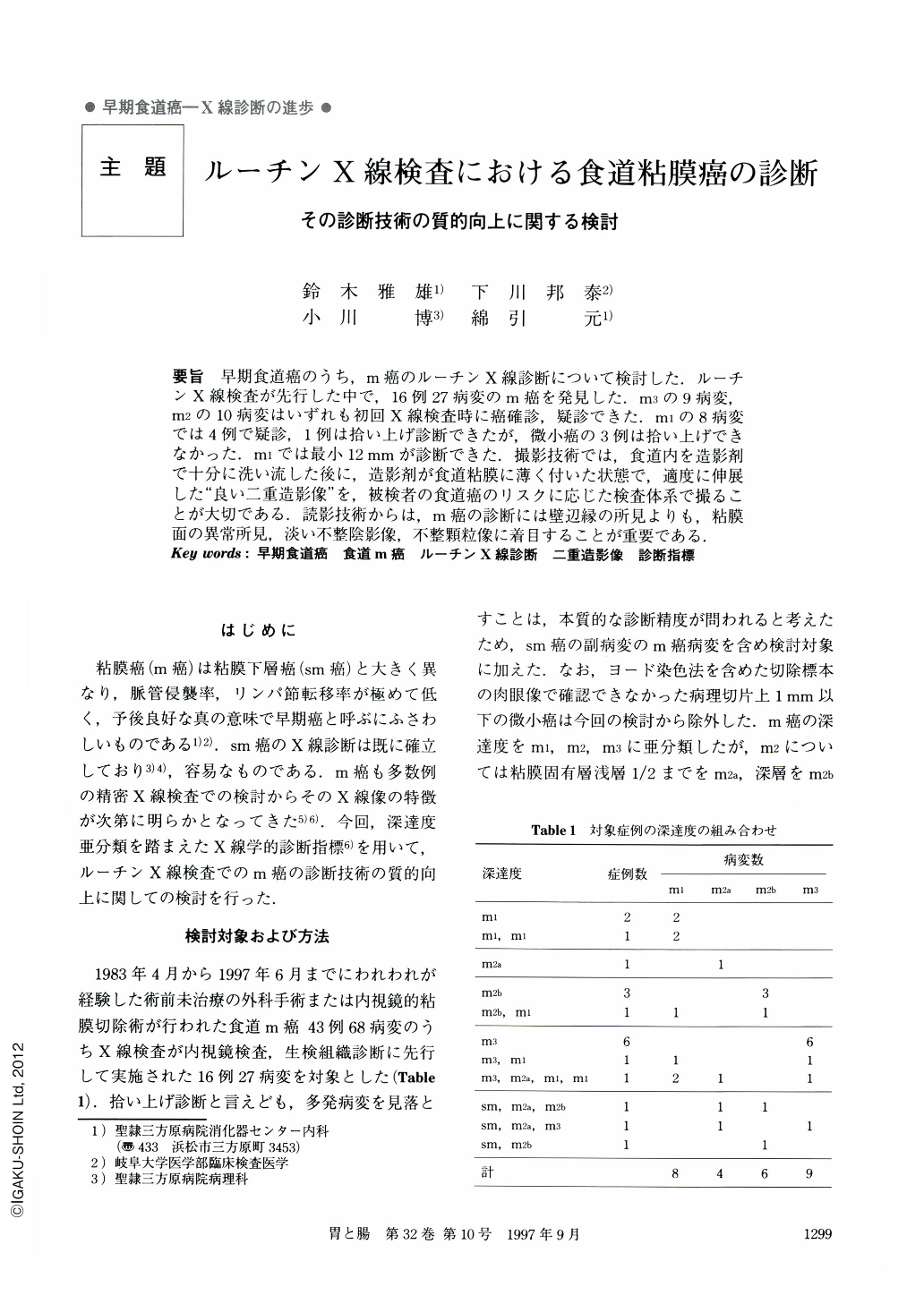
要旨 早期食道癌のうち,m癌のルーチンX線診断について検討した.ルーチンX線検査が先行した中で,16例27病変のm癌を発見した.m3の9病変,m2の10病変はいずれも初回X線検査時に癌確診,疑診できた.m1の8病変では4例で疑診,1例は拾い上げ診断できたが,微小癌の3例は拾い上げできなかった.m1では最小12mmが診断できた.撮影技術では,食道内を造影剤で十分に洗い流した後に,造影剤が食道粘膜に薄く付いた状態で,適度に伸展した“良い二重造影像”を,被検者の食道癌のリスクに応じた検査体系で撮ることが大切である.読影技術からは,m癌の診断には壁辺縁の所見よりも,粘膜面の異常所見,淡い不整陰影像,不整顆粒像に着目することが重要である.
Routine X-ray examination was made in 16 cases, involving 27 lesions of mucosal carcinomas. On the initial X-ray examination, nine lesions were highly suspected of being m3-carcinomas. Ten lesions were thought to the m2-carcinomas. Four lesions were regarded as most probably being m1-carcinomas, while one lesion was diagnosed defintely as being m1-carcinoma. However, three minute carcinomas were missed. In routine examination the smallest m1-carcinoma that can be detected is 12 mm in maximum diameter.
Considering the risk to the patient arising from esophageal carcinoma, it is important to employ a suitable examination process. An X-ray technique using good double-contrast images should be used after washing out the barium in the esophagus, so that it adheres to the esophageal mucosa in a very thin layer suitably. Spread over the mucosal surface. Interpreting the images obtained in the X-ray, it is necessary, when attenpting to diagnose m-carcinomas, not just to pay attention the periphery of the esophageal wall but also to notice any unusual characteristics in the mucosal surface such as faint irregular shadows, or unusual granular quality.
Copyright © 1997, Igaku-Shoin Ltd. All rights reserved.