Japanese
English
- 有料閲覧
- Abstract 文献概要
- 1ページ目 Look Inside
胃静脈瘤に関する報告は従来少なく,食道静脈瘤に比して極めて稀な疾患であると考えられてきた.しかしながら食道静脈瘤の存在する門脈圧充進症患者のレ線像をよく検討すると,胃静脈瘤が併存する場合が稀でないこと,また食道静脈瘤を伴わず胃静脈瘤のみ存在する例もあることが認められてきた1).一方血管外科の発達してきた今日,胃静脈瘤の外科的処理の観点から術前に,病変を正確に把握することは緊急の問題であり,その第一段階として胃静脈瘤の正確な診断が要求されるわけである.最近,食道静脈瘤を伴わず,典型的な胃レ線および内視鏡所見を呈し,経動脈性の門脈造影により確診しえた胃静脈瘤の1例を経験したので報告する.
This is a case of varices of the stomach recently encountered which were unassociatecl with those of the esophagus. They had typical gastric x-ray and endoscopic findings, later accurately diagnosed as such by portal venography.
The patient: a 48-year-old female.
Chief complaint: tarry stool.
Present history: She had been hospitalized for half a year since August, 1966, because liver cirrhosis had been suspected. In the begining of April, 1968, she had a bout of tarry stool, so that she was admitted to the authors' hospital for thorough check-up.
Findings at admission.: Anemia (+), with Hb 37%, jaundice (-) edema (-), aseites (-), vascular spiders (+). The liver, rather firm, was palpated about 2 cm below the eostal margin. Liver function tests.: ieterus index 4, total protein 5.3 g/dl, A/G ratio 0.67, BSP 20%, Kunkel 11 u., GPT 25 u., Alk-P-ase 6u.
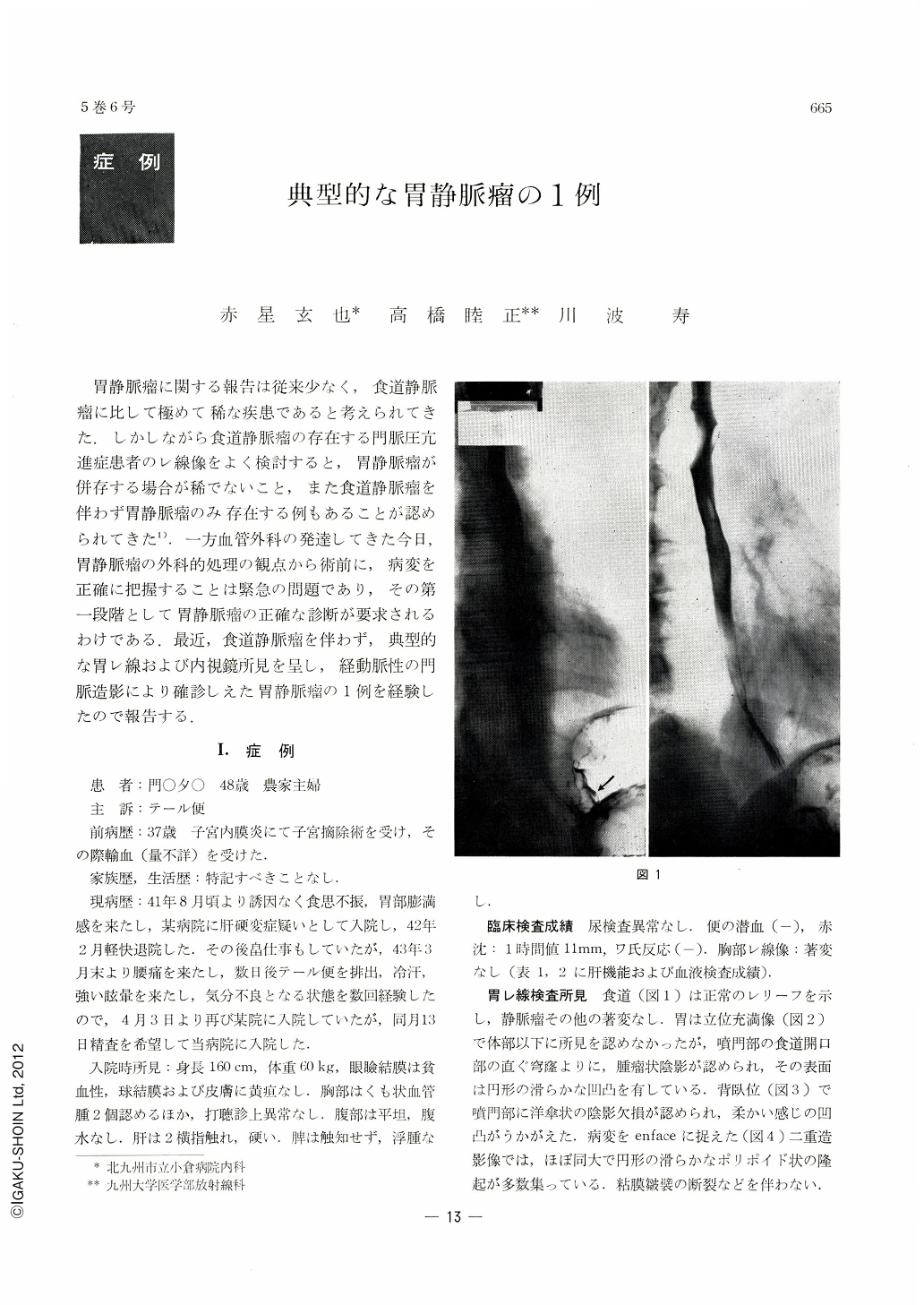
X-ray findings: The esophagus was normal. Tumor-like shadows were noted in the cardia. They were visualized as a group of round, polypoid protrusions, of smooth surface and similar size.
Gastrocamera findings: In the fornix above the cardiac orifice were seen markedly winding protrusions of smooth surface and seemingly of sonsi stency. Neither erosion, bleeding nor reddening was recognized on their surface. These findings clearly indicated that they were gastric varices. For further confirmation of the diagnosis, portal venography via abdominal arteries was conducted. Corresponding to the site of tumors in the cardia, the v. coronaria ventsiculi was seen to have enlarged and become serpentine. The tumor-like protrusions were thus diagnosed as varices. In the past endoscopic diagnosis has been considered difficulut and involving some risk, and only a few cases have been reparted sofar. The authors are, however, of opinion that, as Sakita has asserted, the endoscopic diagnosis of varices of the stomach when unassociated with those of the esophagus hrabors little difficulty if it is done carefully under fluoro scopy.
When general and minute finding together with the site of occurrence are summarizecl, the gastric varices are so characteristic that the diagnosis can be arrived at with accuracy.

Copyright © 1970, Igaku-Shoin Ltd. All rights reserved.


