Japanese
English
- 有料閲覧
- Abstract 文献概要
- 1ページ目 Look Inside
Ⅰ.症例
患者:60歳 ♀
主訴:上腹部膨満感
既往歴,家族歴:特記する事項を認めない.
This is a case of early gastric cancer of type Ⅰ in the cardiac region detected by a gastric mass survey.
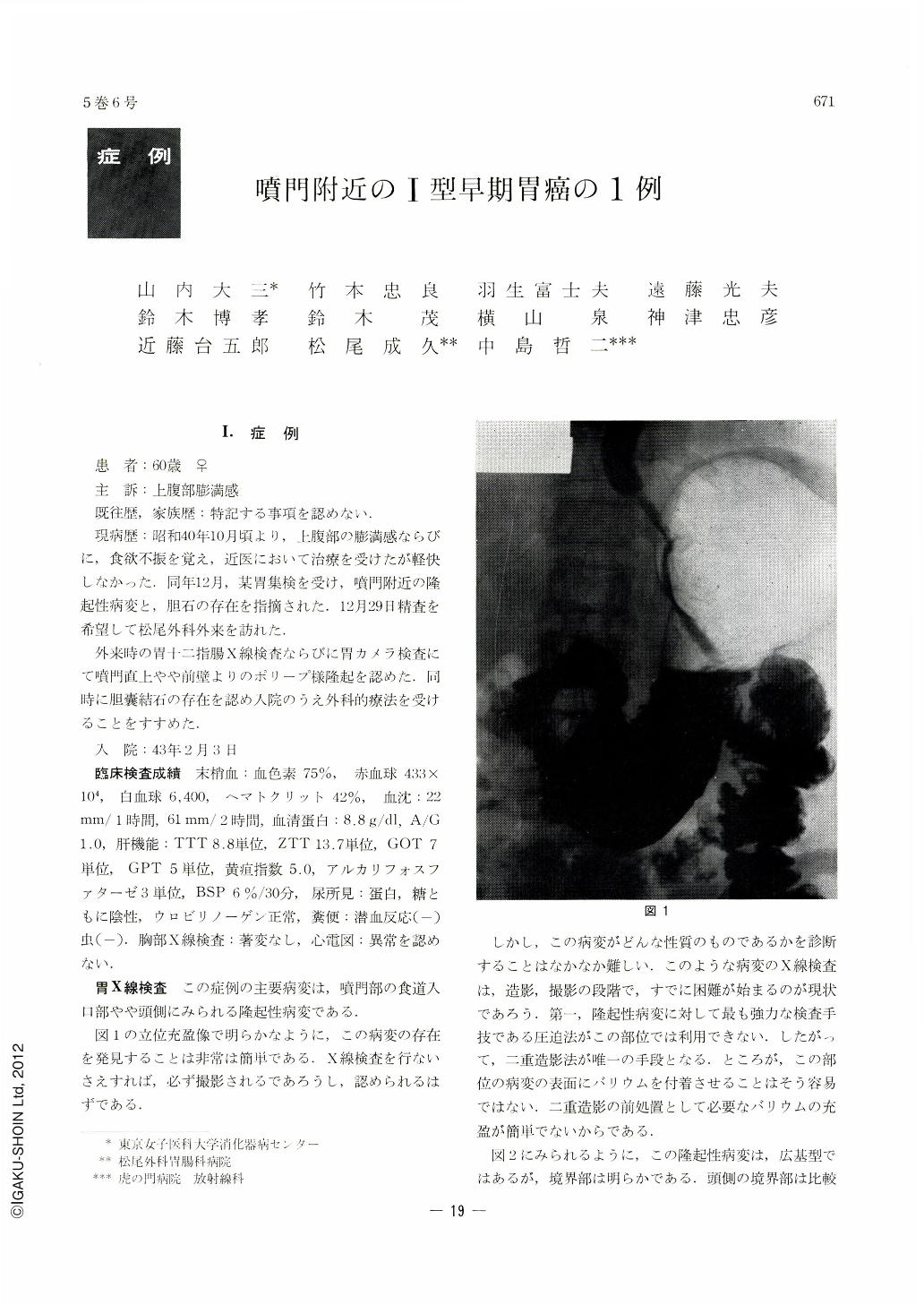
The existence of such an elevated lesion can be proved easily when x-ray gastric pictures are taken in an upright, anterior view, but its qualitative diagnosis is not so easy. In this case gastric x-ray showed an elevated lesion with irregular and granular surface forming two, separated summits, clearly differentiated from the surrounding mucosa, so that either Ⅱa type early cancer or atypical epithelium was suspected.
Endoscopic examination was done with various kinds of instrument so as to get the precise information of this lesion. Va and Vb type of gastric camera were used to see through the wide-angle lens the relatis lesion and the surrounding mucosa.
Dynamic observation and approached vision of this lesion afforded by FGS-C and rigid esophagoscope indicated diversified pictures of surface erosion. Decisive diagnosis was arrived at by obtaining malignant cells among the specimens of sniping biopsy done by FGS-BL using the technic of Ushape retrovision. Histopathological diagnosis after surgery was early gastric cancer of type Ⅰ.
It is of great significance that type Ⅰ early gastric cancer, located in the cardia and therefore very diflicult for discrimination, has been exactly diagnosed before operation.

Copyright © 1970, Igaku-Shoin Ltd. All rights reserved.


