Japanese
English
- 有料閲覧
- Abstract 文献概要
- 1ページ目 Look Inside
Ⅰ.上部消化管大量出血の原因としての門脈圧充進症
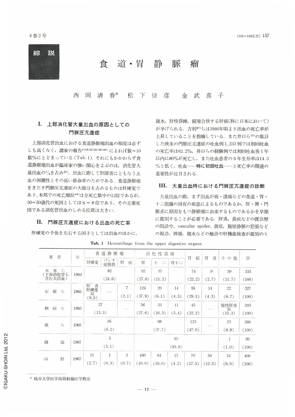
上部消化管出血における食道静脈瘤出血の頻度は必ずしも高くなく,諸家の報告1)19)23)25)26)40)によれば数~10数%にとどまっている(Tab. 1).それにもかかわらず食道静脈瘤出血が臨床家の強い関心をよぶのは,消化管大量出血の1/4を占め23),出血に際して肝障害にともなう止血の困難性とその高い致命率のためである.食道静脈瘤をきたす門脈圧充進症の大部分を占めるものは肝硬変であり,本邦での死亡順位48)は全死亡数中の12位であるが,30~50歳代の死因としては6~8位であり,その主要死因である消化管出血のしめる位置は大きい.
In Japan, bleeding from esophageal varices, usually causing massive hemorrhage, occupies one fourth of massive bleeding from the upper digestive organs. Generally, it is well known that the rupture of esophageal varices plays a leading role in bleeding of portal hypertension, but gastric varices which precede esophageal varices have been of little interest. With respect to the site of obstruction in the portal system the gastric varices are dominant or rarely appear solitarily. To emphasize this point the present study was carried out with barium examination in 62 patients with portal hypertension in whom 10 patients revealed gastric varices only.
The results thus obtained were as follows:
1) Gross findings on the barium roentgenograms were appearance of phrenic ampulla, an increase in the distance between the phrenicus-cardia and between phrenicus-fornix, irregularities of the left hemi-diaphragm, double contour of the lesser curvature and splenomegalia.
2) Fine findings of gastric varices were semi-circular, spindle- and cord-like in shape, which could be in general divided into 3 types, namely, tumor type, polypoid type and socalled giant rugae type, and most of which changed to the giant rugae type with tumor, usually observed in its center. The height of varices was relatively low in most of the cases. The individual margin of the protrusion was smooth, but not sharply defined. Gastric varices mostly appeared around the cardia and then in the fornix, lesser and greater curvature.
3) In most of the patients there were plenty of varices forming a mass in large area of the stomach as mentioned above, but in 3 of 63 patients varices were recognized as a solitary or polyalveoral protrusion which had no relation to the carclia. The most pathognomonic features were alteration the size and shape of the tumor by changing the position of a patient and amount of air in the stomach during the examination. From our results it is necessary to obtain en-face finding of the cardia by the use of double contrast method in the diagnosis of gastric varices.
Copyright © 1969, Igaku-Shoin Ltd. All rights reserved.