Japanese
English
- 有料閲覧
- Abstract 文献概要
- 1ページ目 Look Inside
Ⅰ.症例
患者:須○ト○ 59歳 女子 無職
既往歴:昭和33年虫垂切除術をうけた.
家族歴:特記すべき事項なく,悪性腫瘍の血縁者もいない.
現病歴:昭和42年8月頃空腹時上腹部痛を感じ,某医に胃X線検査をうけたが,特に異常を指摘されなかった.約1カ月間服薬して腹痛は軽快した.昭和43年1月地域胃集団検診において異常所見ありと判定された.
4月9目当院に受診,外来におけるX線検査は不十分であったが,入院のうえ精密検査をすることとし5月1日入院,諸検査の結果早期胃癌と診断し,5月15日胃切除術を施行した症例である.
A 59-year-old female, in whose stomach an abnormal finding had been detected by a local gastric mass survey, was determined as harboring early gastric cancer by a subsequent thorough examination.
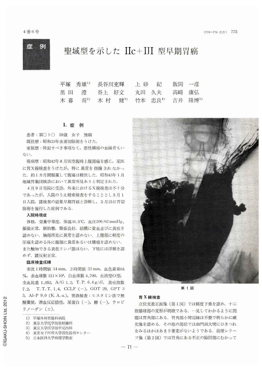
X-ray diagnosis of the stomach: Marked convergence of several mucosal folds was seen in an area ranging from the lesser curvature at the gastric angle away to the posterior wall. Some of these rugae were observed to taper away towards the center, while some others became club-like in their tips. A depressed lesion of irregular shape as well as a nodular protrusion, seen to best advantage in double contrast study, was noted in the center of this area. There were clear double contours in the surrounding region. A shadow suggestive of an ulcer lesion was also noted in the lesser curvature side of these lesions.
Endoscopic diagnosis. An erosion having oval-shaped extent, a little less elevated than the surrounding mucosa, was located at the incisura a little toward the posterior wall. In the center of this depression, a small, shallow ulcer covered over with thick, white exudate was seen. Its closer scrutiny revealed several reddened islet-like protrusions in the depressed region. The mucosal folds, club-like in their tips, abruptly ceased at the edge of the depression. Biopsy was tried on five different spots in this area: they were all positive for cancer.
Pathological diagnosis: A depressed lesion measuring 30 by 10mm was found in the gastric angle a little toward the posterior wall with a very small Ul-III in its center. The latter was encircled all around by ulcer scars having an extent of 18 by 7mm, all covered by regenerated epithelia. These ulcer scars were further surrounded by IIc type depressions with overlying mucosal cancer cells. In short, the cancer lesions had a ring-like extentiona typical holy place type IIc+III early gastric cancer.
An inquiry has been made in this paper about this interesting case as it was further associated with duodenal ulcer.

Copyright © 1969, Igaku-Shoin Ltd. All rights reserved.


