Japanese
English
- 有料閲覧
- Abstract 文献概要
- 1ページ目 Look Inside
Ⅰ.まえがき
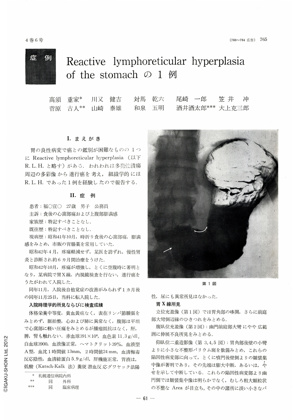
胃の良性病変で癌との鑑別が困難なものの1つにReactive lymphoreticular hyperplasia(以下R. L. H.と略す)がある.われわれは多発仕潰瘍周辺の多彩像から進行癌を考え,組織学的にはR. L. H.であった1例を経験したので報告する.
A case of RLH of the stomach suspected as of Ⅱc type lesion is reported with retrospective analysis of its preoperative clinical diagnosis.
A man 27 years of age, complaining of postcibal epigastralgia and fullness of the upper abdomen, was one year later diagnosed as gastric ulcer by x-ray examination. Abrupt cessation of mucosal folds converging toward the ulcer lesion with their knobby tips was such that IIc type early gastric cancer could not be ruled out. Endoscopically, a depressed lesion, extending from the anterior to posterior wall around the lesser curvature of the pyloric antrum, presented a variegated picture: the depressed area was dotted with multiple small ulcer lesions and erosions with engorgement and yellowish white exudate on its surface. One of the rugae on the anterior wall converging toward the depression showed club-like thickness of its tip, partly altered as if worm-eaten. Another two rugae were fused into one, thickened and discontinued in its way.
Mucosal folds on the posterior wall also were seen to taper off in a stair-like fashion, eventually to merge into the depressed area. The overall picture of the depressed lesion with its rigid appearance was so suspicious of cancer that surgical operation was performed.
Gross observation of the resected stomach showed that mucosal depression, extending from the anterior wall all the way to posterior wall, sharply demarcated all around against the surround ing area, was dotted with many ulcer scars and erosions. This finding was after all very hard to be discriminated from IIc type early cancer. Histopathologically, however, the depressed area previously suspected as of IIc, revealed no cancer cells, but was determined as resultant from pro liferation of lymphatic tissues extensively infiltrating the lamina propria as well as the submucosal layer. Every proliferated lymph follicle was isolated from one another. As there was no atypicality in lymphocytes and reticular cells within it, this case was diagnosed as RLH of the stomach, with reactive proliferation accounting for this lesion.
Copyright © 1969, Igaku-Shoin Ltd. All rights reserved.