Japanese
English
- 有料閲覧
- Abstract 文献概要
- 1ページ目 Look Inside
Ⅰ.はじめに
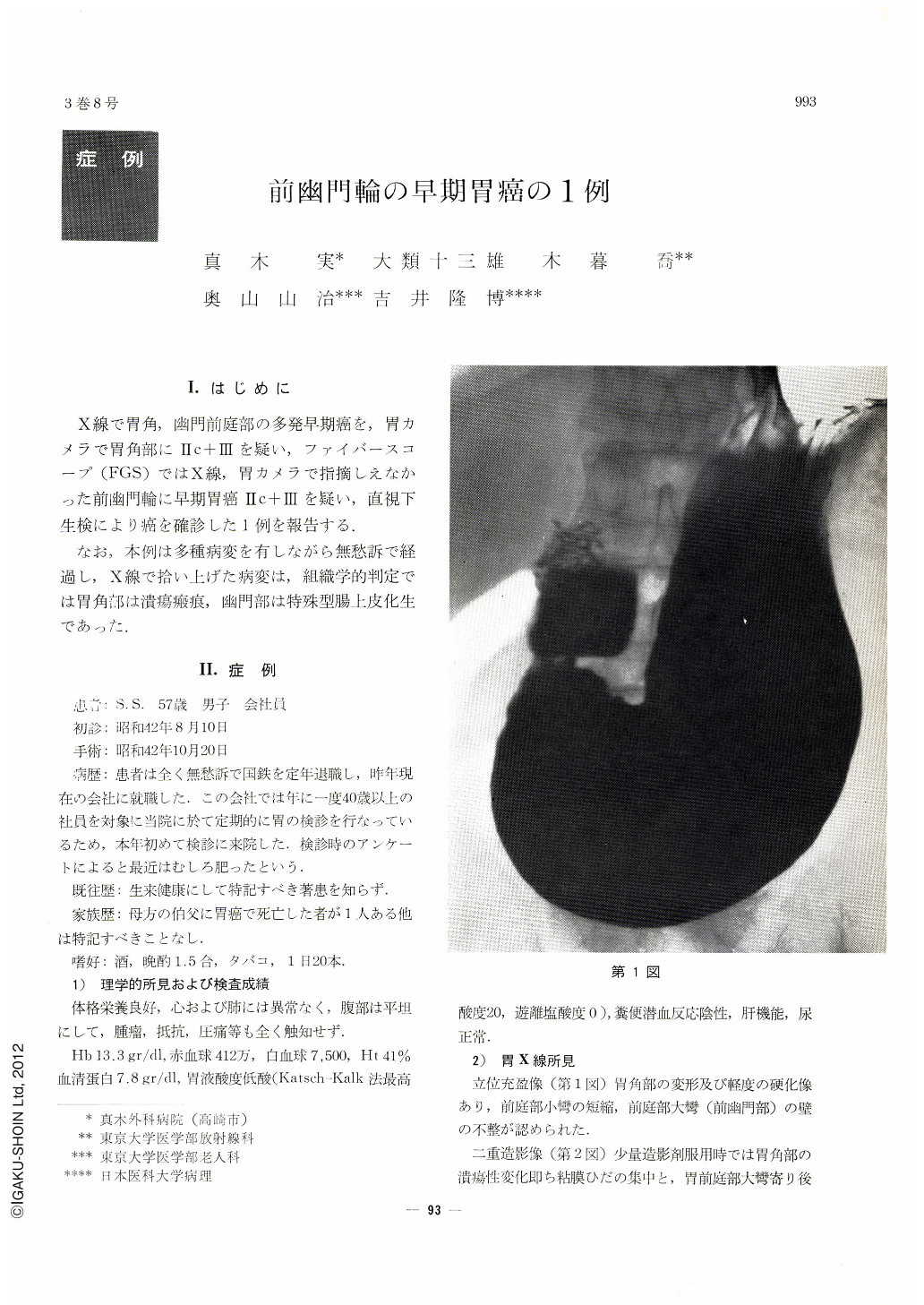
X線で胃角,幽門前庭部の多発早期癌を,胃カメラで胃角部にⅡc+Ⅲを疑い,ファイバースコープ(FGS)ではX線,胃カメラで指摘しえなかった前幽門輪に早期胃癌Ⅱc+Ⅲを疑い,直視下生検により癌を確診した1例を報告する.
なお,本例は多種病変を有しながら無愁訴で経過し,X線で拾い上げた病変は,組織学的判定では胃角部は潰瘍瘢痕,幽門部は特殊型腸上皮化生であった.
One case of early gastric cancer, (Ⅱc+Ⅲ) 1.3×0.5 in size at pre pyloric ring, which produced different results respectively by roentogenological, gastrocamera and fiberscope (F. G. S.) examinations was presented. Histopathological study was sufficiently made. Changes at angulus which was thought to be Ⅱc+Ⅲ by X-ray examination was Ⅱc like scar by F. G. S. examination. Cancer was denied by biopsy. Histopathologically, the change proved to be a healed ulcer. The change at the posterior wall of antrum which had been thought to be Ⅱa+Ⅱc was diagnosed as special type metaplasic gastritis. No malignant change was recognized neither by biopsy nor histopathological examinations. This was also diagnosed as metaplasic gastritis. Some irregularity of the wall of greater curvature was noticed at the oral side of pylorus appeared on a filling image at standing position, but no disease could be pointed out. By F. G. S. examination, there was a doubt of an early cancer at the part of ulcer like change near pylorus ring, reddened excavation at anterior wall nearpylorus and at breaking point of a gastric fold.
Cancer cell was proved by the biopsy under direct view. This case proved the usefulness of F. G. S.-type B to diagnose cancer by biopsy.

Copyright © 1968, Igaku-Shoin Ltd. All rights reserved.


