Japanese
English
- 有料閲覧
PPV購入案内
1,320円 (1,200円+税10%) こちらは、463ページから467ページの文献です。購入には医書.jp本会員登録が必要です。
会員登録完了後に改めて上記「購入サイトへ」を選択してください。
または「購入サイトへ」選択後に「購入ログイン」、「新規会員登録」の順に進んでください。
会員登録について詳しくはをご確認ください。
- Abstract 文献概要
- 1ページ目 Look Inside
症例
患者:52歳 男
主訴:上腹部膨満感
既往歴:42歳のとき急性虫垂炎で虫垂切除ならびに右鼠蹊ヘルニヤの手術をうけている.
家族歴:父が胃癌で死亡している.
A male 52 years of age visited our clinic with a complaint of epigastric fullness.
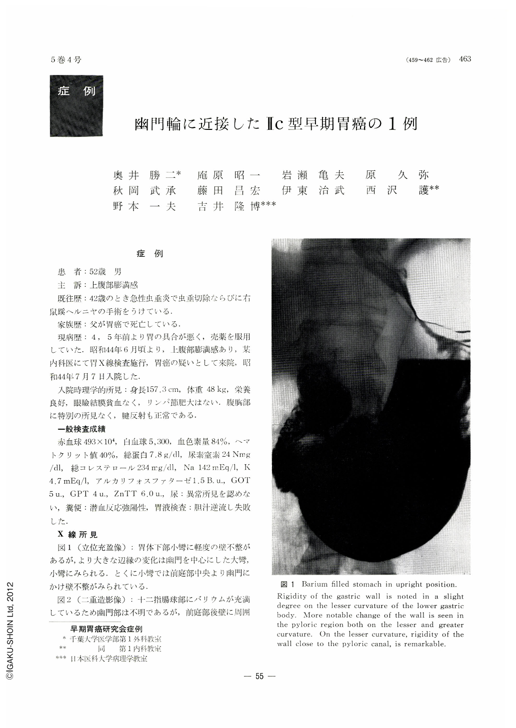
X-ray and endoscopic examinations of the stomach were performed. Barium filled picture of upright frontal view showed stenosis and irregularity of the wall in the pylorus. Double contrast radiograph revealed irregularly granular shadow defects and pale shadow flecks on the posterior wall of the antrum.
Shallow ulcer was detected in prone compression picture. Reddish appearance and depressed, discolored area were discovered by endoscope on the anterior and posterior walls adjacent to the pylorus ring and diagnosed as Ⅱc type. Gastric resection was performed on June 16, 1969.
Operative findings indicated depressed changes on the anterior and posterior walls adjacent to the pylorus ring, mainly localized in the lesser curvature side. No metastasis of lymph nodes was detected. Smear preparation taken during operation showed PAP. class Ⅴ. No cancerous infiltration was confirmed on the anal stump by cytological examination. Gross picture of the resected specimen showed shallow erosion measuring 4.5 by 3cms. Histologically it proved to be adenocarcinoma tubulare, limited to the submucosal layer.
Copyright © 1970, Igaku-Shoin Ltd. All rights reserved.