Japanese
English
- 有料閲覧
- Abstract 文献概要
- 1ページ目 Look Inside
Acanthosis nigricans(黒色表皮腫)は皮膚の乳頭状増殖,色素沈着,角化増生を三徴とし,悪性型(Malignant acanthosis nigricans, MAN)では悪性腫瘍,とりわけ胃癌に併発するため,消化器を扱う者は熟知すべき疾患である.本症は皮膚のみならず,口唇,口腔,食道,直腸,胃,膣などの粘膜をおかすことも知られている.
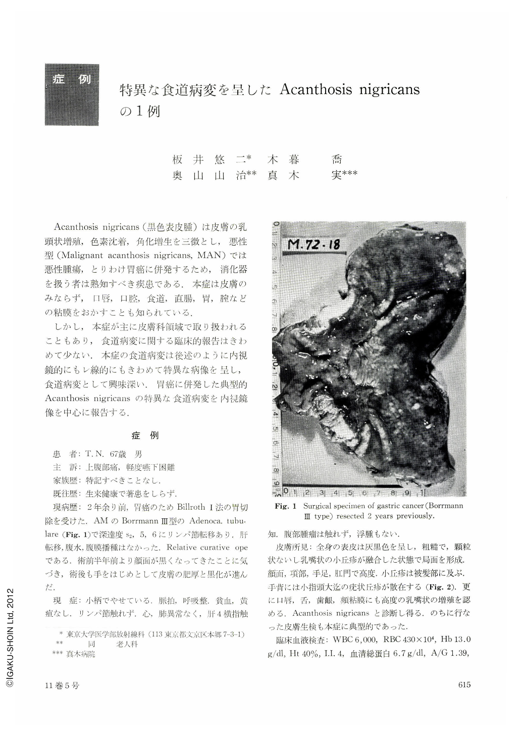
しかし,本症が主に皮膚科領域で取り扱オれることもあり,食道病変に関する臨床的報告はきわめて少ない.本症の食道病変は後述のように内視鏡的にもレ線的にもきわめて特異な病像を呈し,食道病変として興味深い.胃癌に併発した典型的Acanthosis nigricansの特異な食道病変を内視鏡像を中心に報告する.
A 67 year old male patient with history of gastric cancer resected two years previously was admitted with gastric complaints and slight dysphagia. The recurrence of gastric cancer was confirmed by the endoscopical biopsy from the erosion at the gastroduodenal stoma. Barium meal study failed to demonstrate the presence of recurrent cancer but revealed many granular shadows throughout the esophagus. Endoscopically, esophageal mucosa appeared thickened and finely granulated along the entire length of the esophagus as though spawn of fish had been scattered densely. There were several larger elevations up to one centimeter in diameter and in such variation of shapes as discoid, dumbbell-like, worm-like, pyramidal and mimicking a cluster of banana. These lesions were not found beyond the esophago-gastric junction. Endoscopical biopsy specimen of the esophagus showed papillomatous arrangement and widening of the prickle cell layer.
Skin changes were noted almost over the body. Papillomatous and verrucous protrusions as well as hyperpigmentation were intermingled in the face, neck, hands, feet, and perineum. The lips, tongue and oral mucoua were also involved with papillomatous thickening and elevation.
The patient died of recurred cancer four months later despite anticancer chemotherapy.
The esophagus is similar to the skin in being covered by squamous epithelium. Involvement of esophageal mucous membrane in acanthosis nigricans as well as in pemphigoid has been well known among pathologists and dermatologists. However, only a few clinical reports on esophageal manifestation are seen.
Radiological differential diagnosis is necessary from unusual esophageal disorders showing granular shadows such as esophageal moniliasis and progressive systemic sclerosis. Endoscopical diagnosis seems to be easy if the presence of acanthosis nigricans is noted because of close resemblance between esophageal and skin lesions.
Copyright © 1976, Igaku-Shoin Ltd. All rights reserved.