Japanese
English
- 有料閲覧
- Abstract 文献概要
- 1ページ目 Look Inside
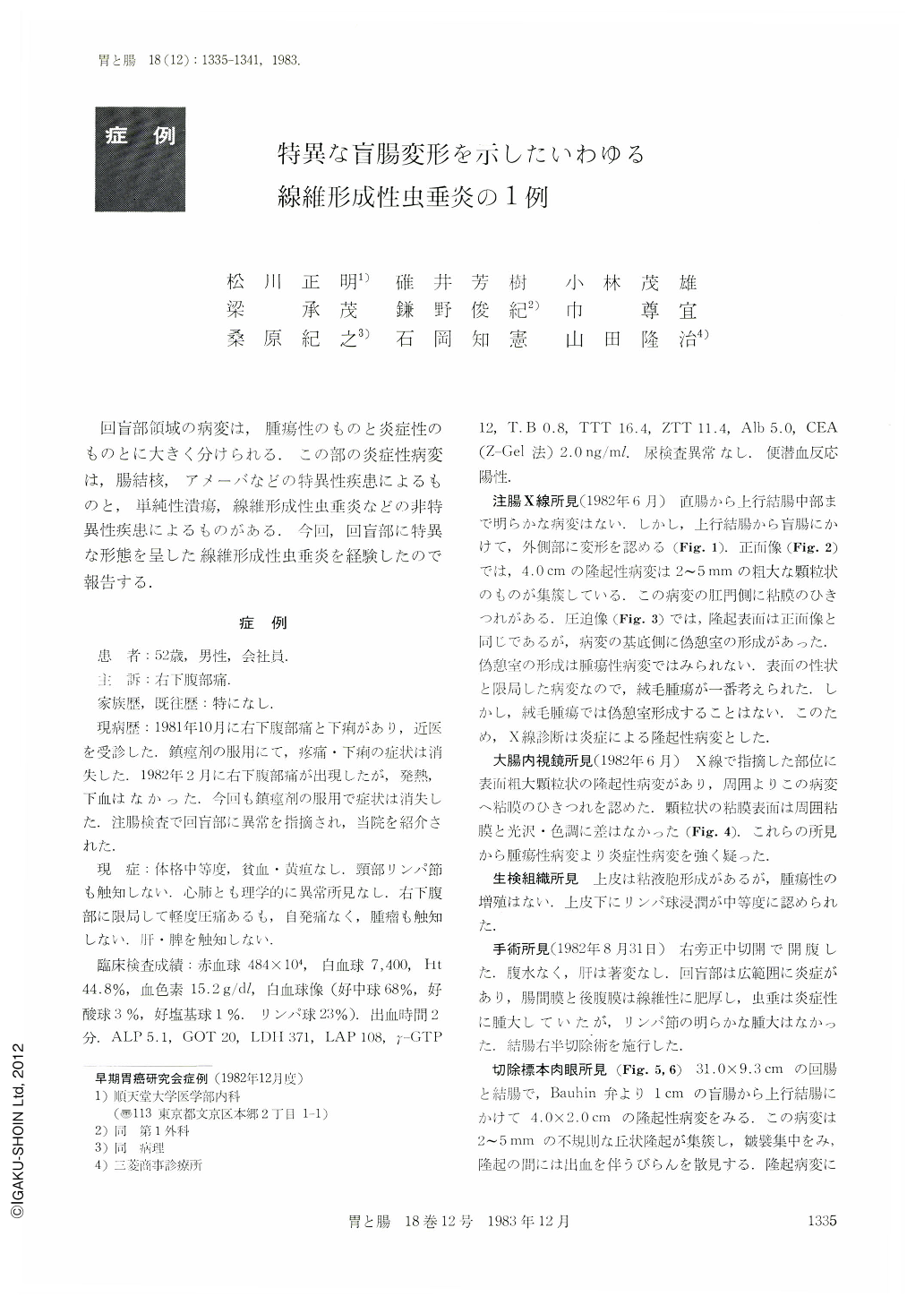
回盲部領域の病変は,腫瘍性のものと炎症性のものとに大きく分けられる.この部の炎症性病変は,腸結核,アメーバなどの特異性疾患によるものと,単純性潰瘍,線維形成性虫垂炎などの非特異性疾患によるものがある.今回,回盲部に特異な形態を呈した線維形成性虫垂炎を経験したので報告する.
症 例
患 者:52歳,男性,会社員.
主 訴:右下腹部痛.
家族歴,既往歴:特になし.
現病歴:1981年10月に右下腹部痛と下痢があり,近医を受診した,鎮痙剤の服用にて,疼痛・下痢の症状は消失した.1982年2月に右下腹部痛が出現したが,発熱,下血はなかった.今回も鎮痙剤の服用で症状は消失した.注腸検査で回盲部に異常を指摘され,当院を紹介された.
A 53-year-old man visited our hospital, complaining of ileocoecal pain twice in six months. His past history was not remarkable. There was no significant family history.
On admission, there was a tender but no palpable tumor in right lower quadrant. A coarse granular lesion and a pseudodiverticular formation at the cecum and proximal portion of the colon ascendens were demonstrated by barium enema.
In colonoscopy, a coarse granular lesion was seen in the region picked up by barium enema.
On surgical exploration, extensive inflammation and fibrous thickening were recognized in ileocoecal region. Right hemicolectomy was carried out.
On resected specimen, a coarse granular lesion (4 cm in diameter) was shown and a longitudinal ulcer scar (2.5 cm in length) distally adjacent to its lesion was recognized. The appendix was swollen to 2 cm in diameter.
In histoiogical examination, chiefly submucosal fibrosis of the appendix and colon, and cystic dilatation of appendicular mucosa with inflammatory changes were recognized. The strongest inflammatory change was at proximal portion of the appendix, but a transmural inflammation, fissuring and fistulation were not recognized. The lesion was diagnosed as socalled fibroplastic appendicitis (Läwen).

Copyright © 1983, Igaku-Shoin Ltd. All rights reserved.


