Japanese
English
- 有料閲覧
- Abstract 文献概要
- 1ページ目 Look Inside
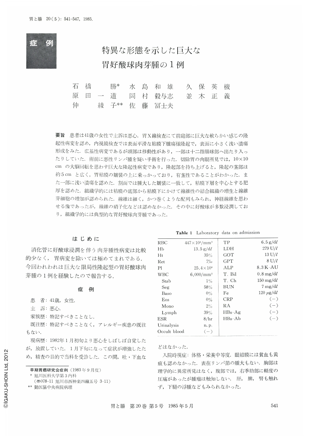
要旨 患者は41歳の女性で主訴は悪心.胃X線検査にて前庭部に巨大な軟らかい感じの隆起性病変を認め,内視鏡検査では表面平滑な粘膜下腫瘍様隆起で,表面に小さく浅い潰瘍形成をみた.広基性病変であるが頭部は移動性があり,一部は十二指腸球部へ出たり入ったりしていた.術前に悪性リンパ腫を疑い手術を行った.切除胃の肉眼所見では,10×10cmの大脳回転を思わす巨大な隆起性病変であり,隆起部を持ち上げると,隆起の茎部は約5cmと広く,胃粘膜の皺襞の上に乗っかっており,有茎性であることがわかった.また一部に浅い潰瘍を認めた.割面では腫大した皺襞に一致して,粘膜下層を中心とする肥厚を認めた.組織学的には粘膜の底部から粘膜下にかけて線維性の結合組織の増生と線維芽細胞の増加が認められた.線維は細く,かつ巻くような配列もみられ,神経線維を思わせる像であったが,線維の硝子化などは認めなかった.その中に好酸球が多数浸潤しており,組織学的には典型的な胃好酸球肉芽腫であった.
The patient was a 41year-old woman with a chief complaint of nausea. Upper gastrointestinal series disclosed a huge protruded lesion in the antrum and endoscopic examination showed a submucosal mass with smooth surface and a small superficial ulceration. The mass had broad base but its head was movable and a part of the tumor was moving in and out between the antrum and the duodenal bulb. She was diagnosed to have malignant lymphoma pre-operatively and an operation was performed. Macroscopic findings of the resected specimen revealed a huge gyrus-like protruded lesion, measuring 10×10cm. The lesion found to be pedunculated when it was lifted-up and a stalk measuring 5 cm was noted on the gastric mucosal fold. Shallow ulceration was also noted partly.
Its cut surface disclosed hypertrophy mainly in the submucosa along the enlarged gastric folds. Histological examination showed proliferation of fibrous connective tissue and proliferation fibroblasts from the mucosal base and the submucosal area. The fibrous tissue had fine fibers with coil arrangement suggesting nerve fibers but hyalinization was not detected. There was massive eosinophilic infiltration in the tissue and histologically, it was a typical case of eosinophilic granuloma of the stomach.

Copyright © 1985, Igaku-Shoin Ltd. All rights reserved.


