Japanese
English
- 有料閲覧
- Abstract 文献概要
- 1ページ目 Look Inside
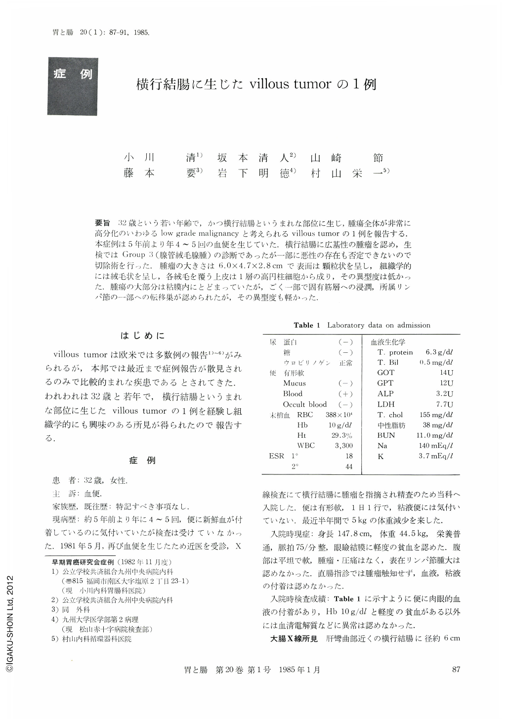
要旨 32歳という若い年齢で,かつ横行結腸というまれな部位に生じ,腫瘍全体が非常に高分化のいわゆるlow grade malignancyと考えられるvillous tumorの1例を報告する.本症例は5年前より年4~5回の血便を生じていた.横行結腸に広基性の腫瘤を認め,生検ではGroup 3(腺管絨毛腺腫)の診断であったが一部に悪性の存在も否定できないので切除術を行った.腫瘤の大きさは6.0×4.7×2.8cmで表面は顆粒状を呈し,組織学的には絨毛状を呈し,各絨毛を覆う上皮は1層の高円柱細胞から成り,その異型度は低かった.腫瘍の大部分は粘膜内にとどまっていたが,ごく一部で固有筋層への浸潤,所属リンパ節の一部への転移巣が認められたが,その異型度も軽かった.
A report is made of a patient as young as 32 years of age, with a villous tumor arising at a place of rare occurance, the transverse colon.
The patient had bouts of bloody stools several times a year since five years ago. Barium enema examination and colonofiberscopy showed a large broad-based tumor of smooth surface in the transverse colon near the hepatic flexure. No formation of ulcer was recognized. Although diagnosis by biopsy was Group 3 (tubullo-villous adenoma), the size of the tumor did not permit us to rule out the existence of malignancy, so that half of the ascending colon and transverse colon was excised with end-to-end anastomosis. In the resected specimen the tumor measured 6.0×4.7×2.8 cm. The surface was granular. Histologically, villouslike projections protruded densely from the muscularis mucosae. The epithelium over each of the villus consisted of a single layer of tall columnar epithelial cells whose nuclei were arranged regularly in the side of the basal membrane. Thus, cellular atypicality was minimal. The majority of the tumor cells remained within the mucous membrane, but in a small part was seen infiltration into the proper muscular layer. Furthermore, a metastatic lesion was seen in the regional lymph node. However, atypicality of tumor cells was slight not only in the infiltrated region but in metastatic nests as well. The present case is thus considered to belong to the so-called adenocarcinoma of low grade malignancy because the tumor itself was highly well differentiated.

Copyright © 1985, Igaku-Shoin Ltd. All rights reserved.


